Human SREC-I/SCARF1 Antibody Summary
Ser20-Thr421
Accession # Q14162
Applications
Please Note: Optimal dilutions should be determined by each laboratory for each application. General Protocols are available in the Technical Information section on our website.
Scientific Data
 View Larger
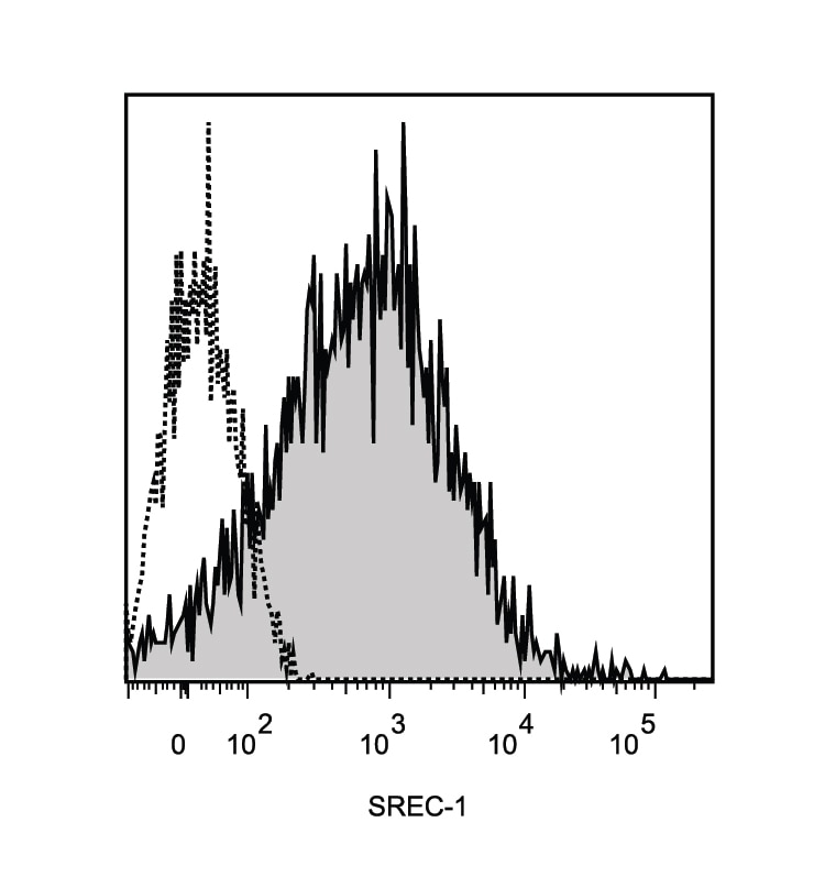
Detection of SREC‑I/SR‑F1 in HUVEC Human Cells by Flow Cytometry. HUVEC human umbilical vein endothelial cells were stained with Goat Anti-Human SREC-I/SR-F1 Antigen Affinity-purified Polyclonal Antibody (Catalog # AF2409, filled histogram) or control antibody (AB-108-C, open histogram), followed by Phycoerythrin-conjugated Anti-Goat IgG Secondary Antibody (F0107).
Reconstitution Calculator
Preparation and Storage
- 12 months from date of receipt, -20 to -70 °C as supplied.
- 1 month, 2 to 8 °C under sterile conditions after reconstitution.
- 6 months, -20 to -70 °C under sterile conditions after reconstitution.
Background: SREC-I/SCARF1
The scavenger receptor (SR) family comprises a group of functionally defined membrane receptors that share a common ability to bind and internalize modified forms of low density lipoproteins (LDL) such as acetylated LDL (AcLDL) and oxidized LDL (OxLDL) (1‑3). Family members are classified alphabetically. They play important roles in lipid metabolism, in host defence and in the regulation of acquired immunity (2, 4). Scavenger receptor expressed by endothelial cells-I (SREC-I) and SREC-II are two proteins that belong to the F type scavenger receptor group (SR-F1 and SR-F2). The full length cDNA of human SREC-I encodes an 830 amino acid (aa) type I transmembrane protein which contains a 19 aa signal peptide, a 402 aa extracellular region, a 21 aa transmembrane segment, and a 388 aa long cytoplasmic domain. The extracellular region contains ten EGF-like repeats (five of which fit the exact consensus sequence for an EGF-like domain) while the cytoplasmic domain is rich in serine and proline in the N-terminal half, and glycine in the C-terminal segment (5, 6). In addition to the full length form, four SREC-I isoforms exist. Two show insertions of a stop codon in EGF-like domain #8, resulting in mature soluble forms of 323 aa and 318 aa, respectively. A third isoform deletes part of domain #8 plus domains #9 and #10; it continues in-frame to generate a mature transmembrane protein of 725 aa. The last isoform shows only cytoplasmic splicing, with 72 aa substituted for the last 332 aa of the full length form. All three transmembrane forms bind acetylated LDL (6). Native SREC-I is approximately 150 kDa in size, and expressed by endothelial cells, macrophages and fetal neurons (7, 8). In the extracellular region human SREC-I is 76% aa identical to mouse SREC-I. The extracellular regions of human SREC-I and II are 53% aa identical.
- Horiuchi, S. et al. (2003) Amino Acids 25:283.
- Greaves, D.R. and S. Gordon (2005) J. Lipid Res. 46:11.
- Platt, N. and S. Gordon (1998) Chem. Biol. 5:R193.
- Platt, N. and S. Gordon (2001) J. Clin. Invest. 108:649.
- Adachi, H. et al. (1997) J. Biol. Chem. 272:31217.
- Adachi, H. and M. Tsujimoto (2002) J. Biol. Chem. 277:24014.
- Shibata, M. et al. (2004) J. Biol. Chem. 279:40084.
- Tanura, Y. et al. (2004) J. Biol. Chem. 279:30938.
Product Datasheets
Citations for Human SREC-I/SCARF1 Antibody
R&D Systems personnel manually curate a database that contains references using R&D Systems products. The data collected includes not only links to publications in PubMed, but also provides information about sample types, species, and experimental conditions.
5
Citations: Showing 1 - 5
Filter your results:
Filter by:
-
A nasal epithelial receptor for Staphylococcus aureus WTA governs adhesion to epithelial cells and modulates nasal colonization.
Authors: Baur S, Rautenberg M, Faulstich M, Grau T, Severin Y, Unger C, Hoffmann W, Rudel T, Autenrieth I, Weidenmaier C
PLoS Pathog, 2014-05-01;10(5):e1004089.
Species: Cotton Rat, Human
Sample Types: Whole Cells
Applications: ICC -
Scavenger receptors in human airway epithelial cells: role in response to double-stranded RNA.
Authors: Dieudonne A, Torres D, Blanchard S, Taront S, Jeannin P, Delneste Y, Pichavant M, Trottein F, Gosset P
PLoS ONE, 2012-08-07;7(8):e41952.
Species: Human
Sample Types: Whole Cells
Applications: Flow Cytometry -
Oligo-guanosine nucleotide induces neuropilin-1 internalization in endothelial cells and inhibits angiogenesis.
Authors: Narazaki M, Segarra M, Hou X, Tanaka T, Li X, Tosato G
Blood, 2010-07-06;116(16):3099-107.
Species: Human
Sample Types: Whole Cells
Applications: Flow Cytometry, ICC -
Sulfated polysaccharides identified as inducers of neuropilin-1 internalization and functional inhibition of VEGF165 and semaphorin3A.
Authors: Narazaki M, Segarra M, Tosato G
Blood, 2008-02-13;111(8):4126-36.
Species: Human
Sample Types: Whole Cells
Applications: Flow Cytometry, ICC -
SCARF1-Induced Efferocytosis Plays an Immunomodulatory Role in Humans, and Autoantibodies Targeting SCARF1 Are Produced in Patients with Systemic Lupus Erythematosus
Authors: April M. Jorge, Taotao Lao, Rachel Kim, Samantha Licciardi, Joseph El Khoury, Andrew D. Luster et al.
The Journal of Immunology
FAQs
No product specific FAQs exist for this product, however you may
View all Antibody FAQsReviews for Human SREC-I/SCARF1 Antibody
Average Rating: 4 (Based on 1 Review)
Have you used Human SREC-I/SCARF1 Antibody?
Submit a review and receive an Amazon gift card.
$25/€18/£15/$25CAN/¥75 Yuan/¥2500 Yen for a review with an image
$10/€7/£6/$10 CAD/¥70 Yuan/¥1110 Yen for a review without an image
Filter by:

