LOX Antibody - BSA Free
Novus Biologicals | Catalog # NB100-2527
![Western Blot: LOX Antibody [NB100-2527]](https://resources.rndsystems.com/images/products/LOX-Antibody-Western-Blot-NB100-2527-img0015.jpg "Western Blot: LOX Antibody [NB100-2527]")
Key Product Details
Validated by
Species Reactivity
Validated:
Cited:
Predicted:
Applications
Validated:
Cited:
Label
Antibody Source
Format
Product Specifications
Immunogen
Reactivity Notes
Localization
Clonality
Host
Isotype
Scientific Data Images for LOX Antibody - BSA Free
![Immunohistochemistry-Paraffin: LOX Antibody [NB100-2527]](https://resources.rndsystems.com/images/products/LOX-Antibody-Immunohistochemistry-Paraffin-NB100-2527-img0017.jpg "Immunohistochemistry-Paraffin: LOX Antibody [NB100-2527]")
Immunohistochemistry-Paraffin: LOX Antibody [NB100-2527]
LOX-Antibody-Immunohistochemistry-Paraffin-NB100-2527-img0017.jpg![Simple Western: LOX Antibody [NB100-2527]](https://resources.rndsystems.com/images/products/LOX-Antibody-Simple-Western-NB100-2527-img0009.jpg "Simple Western: LOX Antibody [NB100-2527]")
Simple Western: LOX Antibody [NB100-2527]
Simple Western: LOX Antibody [NB100-2527] - Simple Western shows a specific band for LOX in 1.0 mg/ml of HeLa cell lysate. This experiment was performed under reducing conditions using the 12-230 kDa separation system.![Immunohistochemistry-Paraffin: LOX Antibody [NB100-2527]](https://resources.rndsystems.com/images/products/LOX-Antibody-Immunohistochemistry-Paraffin-NB100-2527-img0011.jpg "Immunohistochemistry-Paraffin: LOX Antibody [NB100-2527]")
Immunohistochemistry-Paraffin: LOX Antibody [NB100-2527]
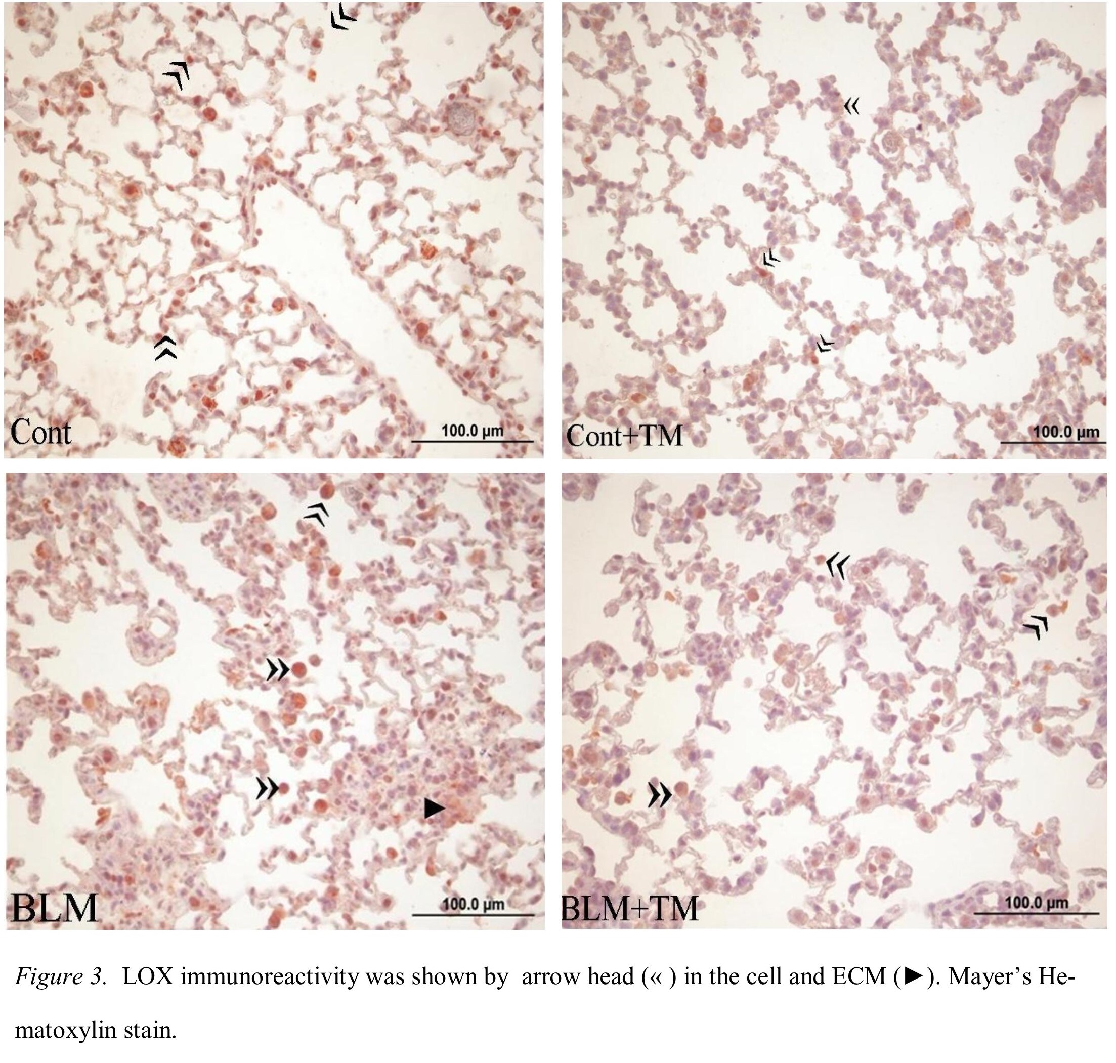
Immunohistochemistry-Paraffin: LOX Antibody [NB100-2527] - LOX in fibrotic mice lung tissue. Primary antibody at 1:100, incubated overnight at 4C. Image from verified customer review.![Knockdown Validated: LOX Antibody [NB100-2527]](https://resources.rndsystems.com/images/products/LOX-Antibody-Knockdown-Validated-NB100-2527-img0016.jpg "Western Blot LOX Antibody [NB100-2527]")
![Immunocytochemistry/ Immunofluorescence: LOX Antibody [NB100-2527]](https://resources.rndsystems.com/images/products/LOX-Antibody-Immunocytochemistry-Immunofluorescence-NB100-2527-img0007.jpg "Immunocytochemistry/ Immunofluorescence: LOX Antibody [NB100-2527]")
Immunocytochemistry/ Immunofluorescence: LOX Antibody [NB100-2527]
Immunocytochemistry/Immunofluorescence: LOX Antibody [NB100-2527] - LOX antibody was tested in HeLa cells with Dylight 488 (green). Nuclei and alpha-tubulin were counterstained with DAPI (blue) and Dylight 550 (red).![Immunohistochemistry: LOX Antibody [NB100-2527]](https://resources.rndsystems.com/images/products/LOX-Antibody-Immunohistochemistry-NB100-2527-img0014.jpg "Immunohistochemistry: LOX Antibody [NB100-2527]")
![Immunocytochemistry/ Immunofluorescence: LOX Antibody [NB100-2527]](https://resources.rndsystems.com/images/products/LOX-Antibody-Immunocytochemistry-Immunofluorescence-NB100-2527-img0013.jpg "Immunocytochemistry/ Immunofluorescence: LOX Antibody [NB100-2527]")
Immunocytochemistry/ Immunofluorescence: LOX Antibody [NB100-2527]
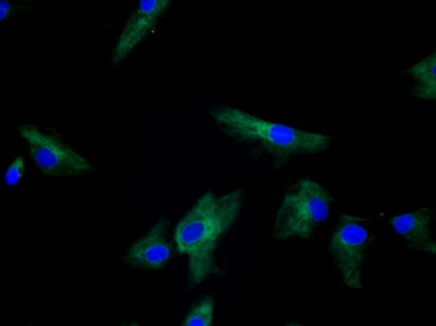
Immunocytochemistry/Immunofluorescence: LOX Antibody [NB100-2527] - Staining in human fibroblasts. Image from verified customer review.![Western Blot: LOX Antibody [NB100-2527]](https://resources.rndsystems.com/images/products/LOX-Antibody-Western-Blot-NB100-2527-img0005.jpg "Western Blot: LOX Antibody [NB100-2527]")
Western Blot: LOX Antibody [NB100-2527]
Western Blot: LOX Antibody [NB100-2527] - Analysis of LOX in human kidney using NB100-2527.![Immunohistochemistry: LOX Antibody [NB100-2527]](https://resources.rndsystems.com/images/products/LOX-Antibody-Immunohistochemistry-NB100-2527-img0006.jpg "Immunohistochemistry: LOX Antibody [NB100-2527]")
Immunohistochemistry: LOX Antibody [NB100-2527]
Immunohistochemistry: LOX Antibody [NB100-2527] - Staining of LOX in mouse stomach.
Western Blot: LOX Antibody [NB100-2527] -
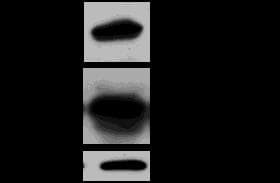
Western Blot: LOX Antibody [NB100-2527] - Patupilone counteracts hypoxia- but not IR-induced LOX-secretion. (A) Western blot of basal & IR-induced LOX in conditioned media derived from patupilone-untreated or patupilone-pretreated (0.5 nM) cells & quantification of band intensities from 3 independent experiments. (B) Western blot of basal & hypoxia-induced LOX in conditioned media derived from patupilone-untreated or patupilone-pretreated (0.5 nM) cells & quantification of band intensities from 3 independent experiments. (C) LOX gene transcription 16 h after irradiation & hypoxia, determined by RT-PCR, averaged over 3 independent experiments. Image collected & cropped by CiteAb from the following publication (https://bmccancer.biomedcentral.com/articles/10.1186/1471-2407-14-532), licensed under a CC-BY license. Not internally tested by Novus Biologicals.Applications for LOX Antibody - BSA Free
Immunocytochemistry/ Immunofluorescence
Immunohistochemistry
Immunohistochemistry-Paraffin
Simple Western
Western Blot
This LOX antibody is useful for Immunocytochemistry/Immunofluorescence, Immunohistochemistry-paraffin embedded sections, and Western blot. In Western blot, bands are observed ~58 kDa representing glycosylated Lox and ~32 kDa representing the mature, secreted form of Lox. In ICC/IF nuclear staining was observed in HeLa cells, which is expected for the mature form of LOX according to published literature (PMID 17287363 and 10996848).
In Simple Western only 10 - 15 uL of the recommended dilution is used per data point.
See Simple Western Antibody Database for Simple Western validation: Tested in HeLa lysate 1.0 mg/mL, separated by Size, antibody dilution of 1:25, apparent MW was 27 kDa. Separated by Size-Wes, Sally Sue/Peggy Sue.
Reviewed Applications
Read 5 reviews rated 4 using NB100-2527 in the following applications:
Formulation, Preparation, and Storage
Purification
Formulation
Format
Preservative
Concentration
Shipping
Stability & Storage
Background: Lysyl Oxidase/LOX
Long Name
Alternate Names
Gene Symbol
Additional Lysyl Oxidase/LOX Products
Product Documents for LOX Antibody - BSA Free
Certificate of Analysis
To download a Certificate of Analysis, please enter a lot or batch number in the search box below.
Product Specific Notices for LOX Antibody - BSA Free
This product is for research use only and is not approved for use in humans or in clinical diagnosis. Primary Antibodies are guaranteed for 1 year from date of receipt.
Citations for LOX Antibody - BSA Free
 Powered by Bioz
Customer Reviews for LOX Antibody - BSA Free (5)
Have you used LOX Antibody - BSA Free?
Submit a review and receive an Amazon gift card!
$25/€18/£15/$25CAN/¥2500 Yen for a review with an image
$10/€7/£6/$10CAN/¥1110 Yen for a review without an image
Submit a review
Customer Images
-
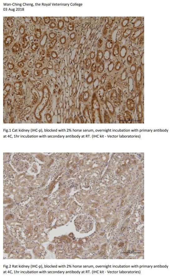
Application: Immunohistochemistry-ParaffinSample Tested: IHC-P Sample TestedSpecies: cat and RatVerified Customer | Posted 08/03/2018

-
Application: ImmunofluorescenceSample Tested: fibroblastsSpecies: HumanVerified Customer | Posted 06/07/2018
-
Application: Immunohistochemistry-ParaffinSample Tested: mouse lungSpecies: MouseVerified Customer | Posted 01/14/2015LOX immunoreactivity in fibrotic mice lung
-
Application: Western BlotSample Tested: mouse vascular smooth muscle cellSpecies: MouseVerified Customer | Posted 03/14/2014LOX protein levels analyzed in cell lysates and supernatants from mice vascular smooth muscle cells
-
Application: Western BlotSample Tested: Renal cell carcinoma cell line UMRC2, Sample Amount: 100ugSpecies: HumanVerified Customer | Posted 06/19/2009
There are no reviews that match your criteria.
Protocols
View specific protocols for LOX Antibody - BSA Free (NB100-2527):
Antigen unmasking
1. Bring slides to a boil in 10 mM sodium citrate buffer pH 6.0 then maintain at a sub-boiling temperature for 10 minutes.
2. Cool slides on bench top for 30 minutes.
Staining
1. Wash sections in dH2O three times for 5 minutes each.
2. Wash section in wash buffer (1X PBS/0.1% Tween-20 (1X PBST)) for 5 minutes.
3. Block each section with 100-400ul blocking solution (1X PBST, 5% goat serum) for 1 hour at room temperature.
4. Remove blocking solution and add 100-400 ul primary antibody diluted in 1X PBST, 5% goat serum to each section.
5. Incubate overnight at 4C.
6. Remove antibody solution and wash sections in wash buffer three times for 5 minutes each.
7. Add 100-400 ul biotinylated secondary antibody, diluted in 1X PBST, 5% goat serum.
8. Incubate 30 minutes at room temperature.
9. Remove secondary antibody solution and wash sections three times with wash buffer for 5 minutes each.
10. Add 100-400 ul Streptavidin HRP reagent to each section and incubate for 30 minutes at room temperature.
11. Wash sections three times in wash buffer for 5 minutes each.
12. Add 100-400 ul DAB substrate to each section and monitor staining closely.
13. As soon as the sections develop, immerse slides in dH2O.
14. Counterstain sections in hematoxylin.
15. Wash sections in dH2O two times for 5 minutes each.
16. Dehydrate sections.
17. Mount coverslips.
1. Perform SDS-PAGE (4-12%, Bis-Tris) on samples to be analyzed, loading 40 ug of total protein per lane.
2. Transfer proteins to Nitrocellulose according to the instructions provided by the manufacturer of the transfer apparatus.
3. Rinse membrane with dH2O and then stain the blot using ponceau S for 1-2 minutes to access the transfer of proteins onto the nitrocellulose membrane. Rinse the blot in water to remove excess stain and mark the lane locations and locations of molecular weight markers using a pencil.
4. Rinse the blot in TBS for approximately 5 minutes.
5. Block the membrane using 5% non-fat dry milk + 1% BSA in TBS, overnight at 4C.
6. Rinse the membrane in dH2O and then wash the membrane in wash buffer [TBS + 0.1% Tween] 3 times for 10 minutes each.
7. Dilute the rabbit anti-LOX primary antibody (NB 100-2527) in blocking buffer and incubate 1 hour at room temperature.
8. Rinse the membrane in dH2O and then wash the membrane in wash buffer [TBS + 0.1% Tween] 3 times for 10 minutes each.
9. Apply the diluted rabbit-IgG HRP-conjugated secondary antibody in blocking buffer (as per manufacturer's instructions) and incubate 1 hour at room temperature.
10. Wash the blot in wash buffer [TBS + 0.1% Tween] 3 times for 10 minutes each (this step can be repeated as required to reduce background).
11. Apply the detection reagent of choice in accordance with the manufacturer's instructions (Pierce's ECL).
**Note: Tween-20 can be added to the blocking or antibody dilution buffer at a final concentration of 0.05-0.2%, provided it does not interfere with antibody-antigen binding.
Find general support by application which include: protocols, troubleshooting, illustrated assays, videos and webinars.
- Antigen Retrieval Protocol (PIER)
- Antigen Retrieval for Frozen Sections Protocol
- Appropriate Fixation of IHC/ICC Samples
- Cellular Response to Hypoxia Protocols
- Chromogenic IHC Staining of Formalin-Fixed Paraffin-Embedded (FFPE) Tissue Protocol
- Chromogenic Immunohistochemistry Staining of Frozen Tissue
- ClariTSA™ Fluorophore Kits
- Detection & Visualization of Antibody Binding
- Fluorescent IHC Staining of Frozen Tissue Protocol
- Graphic Protocol for Heat-induced Epitope Retrieval
- Graphic Protocol for the Preparation and Fluorescent IHC Staining of Frozen Tissue Sections
- Graphic Protocol for the Preparation and Fluorescent IHC Staining of Paraffin-embedded Tissue Sections
- Graphic Protocol for the Preparation of Gelatin-coated Slides for Histological Tissue Sections
- ICC Cell Smear Protocol for Suspension Cells
- ICC Immunocytochemistry Protocol Videos
- ICC for Adherent Cells
- IHC Sample Preparation (Frozen sections vs Paraffin)
- Immunocytochemistry (ICC) Protocol
- Immunocytochemistry Troubleshooting
- Immunofluorescence of Organoids Embedded in Cultrex Basement Membrane Extract
- Immunofluorescent IHC Staining of Formalin-Fixed Paraffin-Embedded (FFPE) Tissue Protocol
- Immunohistochemistry (IHC) and Immunocytochemistry (ICC) Protocols
- Immunohistochemistry Frozen Troubleshooting
- Immunohistochemistry Paraffin Troubleshooting
- Preparing Samples for IHC/ICC Experiments
- Preventing Non-Specific Staining (Non-Specific Binding)
- Primary Antibody Selection & Optimization
- Protocol for Heat-Induced Epitope Retrieval (HIER)
- Protocol for Making a 4% Formaldehyde Solution in PBS
- Protocol for VisUCyte™ HRP Polymer Detection Reagent
- Protocol for the Fluorescent ICC Staining of Cell Smears - Graphic
- Protocol for the Fluorescent ICC Staining of Cultured Cells on Coverslips - Graphic
- Protocol for the Preparation & Fixation of Cells on Coverslips
- Protocol for the Preparation and Chromogenic IHC Staining of Frozen Tissue Sections
- Protocol for the Preparation and Chromogenic IHC Staining of Frozen Tissue Sections - Graphic
- Protocol for the Preparation and Chromogenic IHC Staining of Paraffin-embedded Tissue Sections
- Protocol for the Preparation and Chromogenic IHC Staining of Paraffin-embedded Tissue Sections - Graphic
- Protocol for the Preparation and Fluorescent ICC Staining of Cells on Coverslips
- Protocol for the Preparation and Fluorescent ICC Staining of Non-adherent Cells
- Protocol for the Preparation and Fluorescent ICC Staining of Stem Cells on Coverslips
- Protocol for the Preparation and Fluorescent IHC Staining of Frozen Tissue Sections
- Protocol for the Preparation and Fluorescent IHC Staining of Paraffin-embedded Tissue Sections
- Protocol for the Preparation of Gelatin-coated Slides for Histological Tissue Sections
- Protocol for the Preparation of a Cell Smear for Non-adherent Cell ICC - Graphic
- R&D Systems Quality Control Western Blot Protocol
- TUNEL and Active Caspase-3 Detection by IHC/ICC Protocol
- The Importance of IHC/ICC Controls
- Troubleshooting Guide: Immunohistochemistry
- Troubleshooting Guide: Western Blot Figures
- Western Blot Conditions
- Western Blot Protocol
- Western Blot Protocol for Cell Lysates
- Western Blot Troubleshooting
- Western Blot Troubleshooting Guide
- View all Protocols, Troubleshooting, Illustrated assays and Webinars
FAQs for LOX Antibody - BSA Free
-
Q: Hi, I was wondering if you have any blocking peptides for NB100-59729, NB100-2527 and NB100-2530?
A: Catalog # NB100-2527PEP is the blocking peptide for catalog # NB100-2527.