Mouse CD28 Antibody Summary
Accession # P31041
Applications
Please Note: Optimal dilutions should be determined by each laboratory for each application. General Protocols are available in the Technical Information section on our website.
Scientific Data
 View Larger
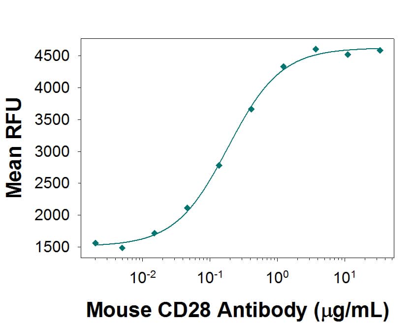
Mouse CD28 Antibody Induces Proliferation in Mouse T Cells. Rat Anti-Mouse CD28 Monoclonal Antibody (Catalog # MAB4832) induces proliferation in mouse T cells in the presence of 100 ng/mL Hamster Anti-Mouse CD3e Monoclonal Antibody (Catalog # MAB484), in a dose dependent manner, as measured by Resazurin (Catalog # AR002), The ED50 for this effect is typically 0.07-0.42 µg/mL.
 View Larger
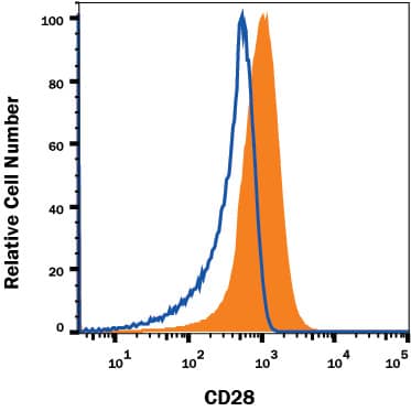
Detection of CD28 in Mouse Thymocytes by Flow Cytometry. Mouse thymocytes were stained with Rat Anti-Mouse CD28 Monoclonal Antibody (Catalog # MAB4832, filled histogram) or isotype control antibody (MAB005, open histogram), followed by Phycoerythrin-conjugated Anti-Rat IgG Secondary Antibody (F0105B). Staining was performed using our Membrane-Associated Proteins protocol.
 View Larger
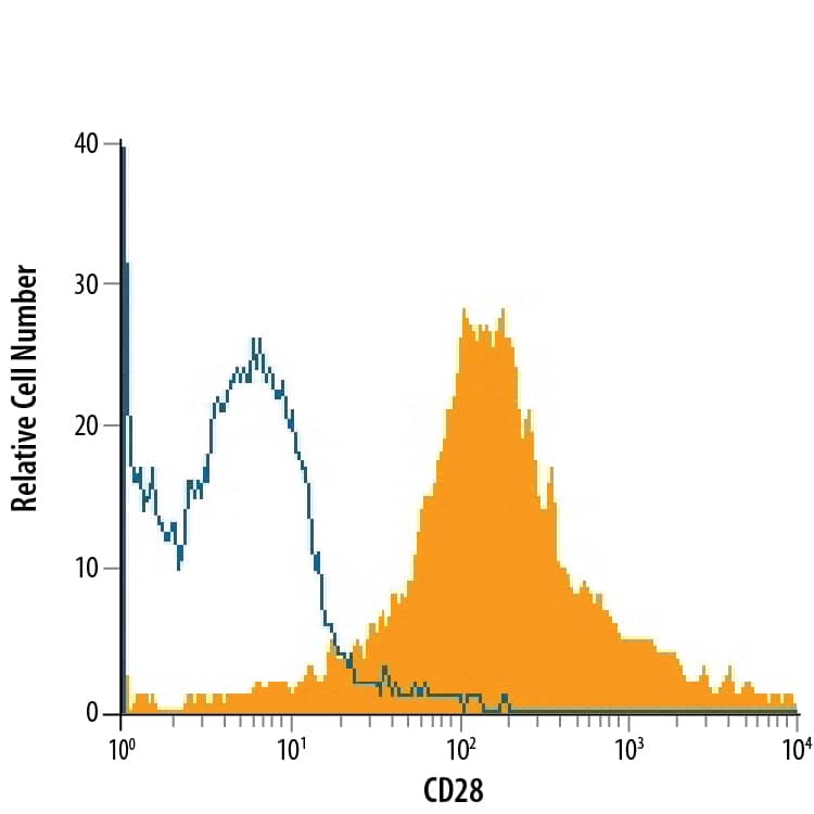
Detection of CD28 in CD3+Mouse Splenocytes by Flow Cytometry. CD3+mouse splenocytes were stained with Rat Anti-Mouse CD28 Monoclonal Antibody (Catalog # MAB4832, filled histogram) or isotype control antibody (Catalog # MAB005, open histogram), followed by Phycoerythrin-conjugated Anti-Rat IgG Secondary Antibody (Catalog # F0105B).
Reconstitution Calculator
Preparation and Storage
- 12 months from date of receipt, -20 to -70 °C as supplied.
- 1 month, 2 to 8 °C under sterile conditions after reconstitution.
- 6 months, -20 to -70 °C under sterile conditions after reconstitution.
Background: CD28
CD28 and CTLA-4, together with their ligands B7-1 and B7-2, constitute one of the dominant costimulatory pathways that regulate T and B cell responses. CD28 and CTLA-4 are structurally homologous molecules that are members of the immunoglobulin (Ig) gene superfamily. Both CD28 and CTLA-4 are composed of a single Ig
V‑like extracellular domain, a transmembrane domain and an intracellular domain. CD28 and CTLA-4 are both expressed on the cell surface as disulfide-linked homodimers or as monomers. The genes encoding these two molecules are closely linked on human chromosome 2 and mouse chromosome 1. Mouse CD28 is expressed constitutively on virtually 100% of mouse T cells and on developing thymocytes. Cell surface expression of mouse CD28 is down-regulated upon ligation of CD28 in the presence of PMA or PHA. In contrast, CTLA-4 is not expressed constitutively but is upregulated rapidly following T cell activation and CD28 ligation. Cell surface expression of CTLA-4 peaks approximately 48 hours after activation. Although both CTLA-4 and CD28 can bind to the same ligands, CTLA-4 binds to
B7‑1 and B7‑2 with a 20-100 fold higher affinity than CD28. CD28/B7 interaction has been shown to prevent apoptosis of activated T cells via the up-regulation of
Bcl‑xL. CD28 ligation has also been shown to regulate Th1/Th2 differentiation. Agonist activity has been reported using MAB4831 (4,5).
- Lenschow, D.J. et al. (1996) Annu. Rev. Immunol. 14:233.
- Hathcock, K.S. and R.J. Hodes (1996) Advances in Immunol. 62:131.
- Ward, S.G. (1996) Biochem. J. 318:361.
- Nguyen, P. et al. (2003) Blood 13:4320.
- Orbach, A. et al. (2007) J. Immunol. 179:7287.
Product Datasheets
Citations for Mouse CD28 Antibody
R&D Systems personnel manually curate a database that contains references using R&D Systems products. The data collected includes not only links to publications in PubMed, but also provides information about sample types, species, and experimental conditions.
6
Citations: Showing 1 - 6
Filter your results:
Filter by:
-
MicroRNA‑30a controls the instability of inducible CD4+ Tregs through SOCS1
Authors: Ya Zhou, Yongju Li, Jia Lu, Xiaowu Hong, Lin Xu
Molecular Medicine Reports
-
The YAP/HIF-1&alpha/miR-182/EGR2 axis is implicated in asthma severity through the control of Th17 cell differentiation
Authors: J Zhou, N Zhang, W Zhang, C Lu, F Xu
Cell & bioscience, 2021-05-12;11(1):84.
Species: Mouse
Sample Types: Whole Cells
Applications: Neutralization -
Crucial role of OX40/OX40L signaling in a murine model of asthma
Authors: W Lei, D Zeng, G Liu, Y Zhu, J Wang, H Wu, J Jiang, J Huang
Mol Med Rep, 2018-01-18;0(0):.
Species: Mouse
Sample Types: Whole Cells
Applications: Functional Assay -
Aloperine Protects Mice against DSS-Induced Colitis by PP2A-Mediated PI3K/Akt/mTOR Signaling Suppression
Authors: X Fu, F Sun, F Wang, J Zhang, B Zheng, J Zhong, T Yue, X Zheng, JF Xu, CY Wang
Mediators Inflamm., 2017-09-19;2017(0):5706152.
Species: Mouse
Sample Types: Whole Cells
Applications: Functional Assay -
The excretory-secretory products of Echinococcus granulosus protoscoleces directly regulate the differentiation of B10, B17 and Th17 cells
Authors: W Pan, WT Hao, YJ Shen, XY Li, YJ Wang, FF Sun, JH Yin, J Zhang, RX Tang, JP Cao, KY Zheng
Parasit Vectors, 2017-07-21;10(1):348.
Species: Mouse
Sample Types: Whole Cells
Applications: Functional Assay -
Temporal Expression of Bim Limits the Development of Agonist-Selected Thymocytes and Skews Their TCR? Repertoire
Authors: David A Hildeman
J. Immunol., 2016-11-16;0(0):.
Species: Mouse
Sample Types: Whole Cells
Applications: Functional Assay
FAQs
No product specific FAQs exist for this product, however you may
View all Antibody FAQsReviews for Mouse CD28 Antibody
There are currently no reviews for this product. Be the first to review Mouse CD28 Antibody and earn rewards!
Have you used Mouse CD28 Antibody?
Submit a review and receive an Amazon gift card.
$25/€18/£15/$25CAN/¥75 Yuan/¥2500 Yen for a review with an image
$10/€7/£6/$10 CAD/¥70 Yuan/¥1110 Yen for a review without an image


