Mouse JAM-C (CD323) APC-conjugated Antibody
Mouse JAM-C (CD323) APC-conjugated Antibody Summary
Val32-Asn241
Accession # Q9D8B7
Applications
Please Note: Optimal dilutions should be determined by each laboratory for each application. General Protocols are available in the Technical Information section on our website.
Scientific Data
 View Larger
Detection of JAM‑C (CD323) in B16‑F1 Mouse Cell Line by Flow Cytometry. B16-F1 mouse melanoma cell line was stained with Rat Anti-Mouse JAM-C (CD323) APC-conjugated Monoclonal Antibody (Catalog # FAB7050A, filled histogram) or isotype control antibody (Catalog # IC013A, open histogram). View our protocol for Staining Membrane-associated Proteins.
Preparation and Storage
Background: JAM-C
JAM-C (Junctional Adhesion Molecule-C), also known as JAM-3 and JAM-2, is a 40-45 kDa member of the JAM family, IgSF of molecules. It is a type I transmembrane glycoprotein that is further classified as a classical JAM with a short cytoplasmic tail vs. non-classical JAMs that contain long cytoplasmic tails. Mature mouse JAM-C is 289 amino acids (aa) in length, and contains a 212 aa extracellular region. This region possesses one N-terminal V-type and one C2-type Ig-like domain (aa 30-241). In human, JAM-C is perhaps best known as a component of the epithelial cell tight junction. In concert with two other transmembrane protein types (claudins and occludin) in-cis, JAM-C forms an intercellular barrier complex that restricts paracellular permeability. In mouse, however, this application may not apply, as endothelium, rather than epithelium, appears to be site of maximum expression. In this locale, JAM-C is suggested to regulate neutrophil exodus from the blood, and provide a barrier against reverse migration back into the vasculature. In any event, binding partners for JAM-C in-trans include JAM-B, Mac-1, alpha x beta 2 and JAM-C itself. Cells known to express JAM-C in mouse do not necessarily mirror those expressing JAM-C in human. Mouse cells known to contain JAM-C are limited in type and include endothelium, high endothelial venules, fibroblasts, hematopoietic stem cells, Schwann cells involved in myelination, and neurons of an Aδ (or pain sensing) phenotype. The extracellular domain of mouse JAM-C shares 96% and 87% aa sequence identity with the extracellular domains of rat and human JAM-C, respectively.
Product Datasheets
Citations for Mouse JAM-C (CD323) APC-conjugated Antibody
R&D Systems personnel manually curate a database that contains references using R&D Systems products. The data collected includes not only links to publications in PubMed, but also provides information about sample types, species, and experimental conditions.
3
Citations: Showing 1 - 3
Filter your results:
Filter by:
-
Neutrophil trogocytosis during their trans-endothelial migration: role of extracellular CIRP
Authors: Satoshi Takizawa, Yongchan Lee, Asha Jacob, Monowar Aziz, Ping Wang
Molecular Medicine
-
Junctional Adhesion Molecule 2 Represents a Subset of Hematopoietic Stem Cells with Enhanced Potential for T Lymphopoiesis
Authors: V Radulovic, M van der Ga, S Koide, V Sigurdsson, S Lang, S Kaneko, K Miharada
Cell Rep, 2019-06-04;27(10):2826-2836.e5.
Species: Mouse
Sample Types: Whole Cells
Applications: Flow Cytometry -
Function of Jam-B/Jam-C interaction in homing and mobilization of human and mouse hematopoietic stem and progenitor cells.
Authors: Arcangeli M, Bardin F, Frontera V, Bidaut G, Obrados E, Adams R, Chabannon C, Aurrand-Lions M
Stem Cells, 2014-04-01;32(4):1043-54.
Species: Human
Sample Types: Whole Cells
Applications: Flow Cytometry
FAQs
No product specific FAQs exist for this product, however you may
View all Antibody FAQsReviews for Mouse JAM-C (CD323) APC-conjugated Antibody
Average Rating: 4 (Based on 1 Review)
Have you used Mouse JAM-C (CD323) APC-conjugated Antibody?
Submit a review and receive an Amazon gift card.
$25/€18/£15/$25CAN/¥75 Yuan/¥2500 Yen for a review with an image
$10/€7/£6/$10 CAD/¥70 Yuan/¥1110 Yen for a review without an image
Filter by:
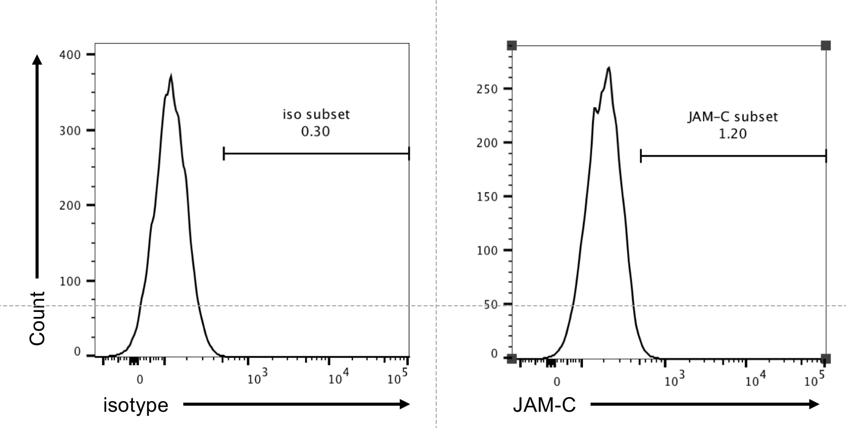
Cells were pre-gated with live/dead and FSC-A/W to exclude dead cells and cell doublets.
The single cell suspension was isolated from lung of a WT B6 mouse and stained with the JAM-C (CD323) APC-conjugated antibody (1:100) for 30 minutes. Cells were analyzed by flow cytometer.


