Mouse PEAR1 Antibody Summary
Leu19-Leu754
Accession # Q8VIK5
Applications
Please Note: Optimal dilutions should be determined by each laboratory for each application. General Protocols are available in the Technical Information section on our website.
Scientific Data
 View Larger
Detection of Mouse PEAR1 by Western Blot. Western blot shows lysates of mouse spleen tissue and mouse platelets. PVDF membrane was probed with 1 µg/mL of Sheep Anti-Mouse PEAR1 Antigen Affinity-purified Polyclonal Antibody (Catalog # AF7607) followed by HRP-conjugated Anti-Sheep IgG Secondary Antibody (Catalog # HAF016). A specific band was detected for PEAR1 at approximately 120-150 kDa (as indicated). This experiment was conducted under reducing conditions and using Immunoblot Buffer Group 1.
 View Larger
Detection of PEAR1 in bEnd.3 Mouse Cell Line by Flow Cytometry. bEnd.3 mouse endo-thelioma cell line was stained with Sheep Anti-Mouse PEAR1 Antigen Affinity-purified Polyclonal Antibody (Catalog # AF7607, filled histogram) or isotype control antibody (Catalog # 5-001-A, open histogram), followed by Phycoerythrin-conjugated Anti-Sheep IgG Secondary Antibody (Catalog # F0126).
 View Larger
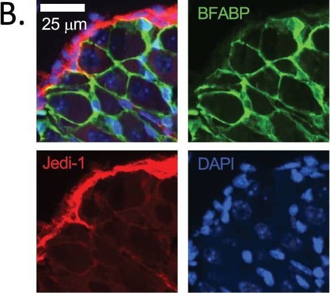
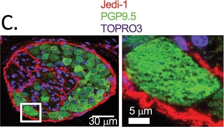
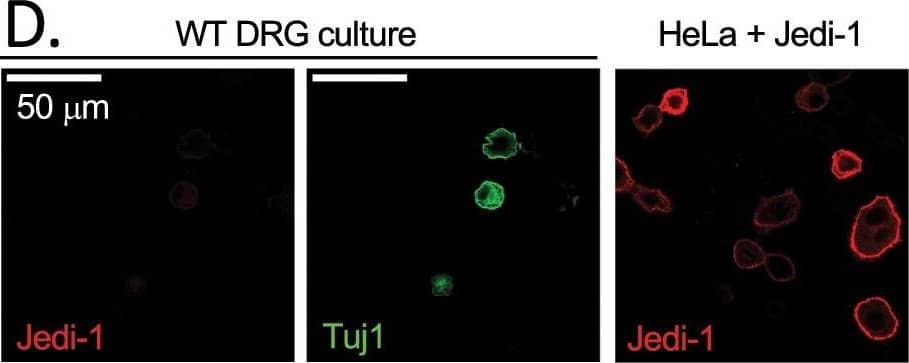
Detection of Mouse PEAR1 by Immunohistochemistry Jedi is expressed in peripheral glia and endothelium. (A) Cross section of adult (8–12 weeks old) WT sciatic nerve stained for Jedi-1 (magenta) and DAPI (blue). Insets 1 and 2 show Jedi expression in blood vessels. See Supplementary Fig. S2 for validation of Jedi-1 expression in perineurial glial cells. (B) WT P0 DRG co-stained for Jedi-1 (red), satellite glial marker BFABP (green), and DAPI (blue). (C) WT DRG co-stained for Jedi-1 (red) and PGP9.5 (green) and TOPRO3 (blue). Right shows inset. (D) Right: Primary WT DRG cultures stained for neurons using Tuj1 (green) and Jedi-1 (red). Left: HeLa cells overexpressing mouse Jedi-1 were used as a positive control for Jedi-1 immunocytochemistry in vitro. All images were analyzed in ImageJ version 2.0.0-rc-69/1.52p. Image collected and cropped by CiteAb from the following publication (https://pubmed.ncbi.nlm.nih.gov/31992767), licensed under a CC-BY license. Not internally tested by R&D Systems.
 View Larger
Detection of Mouse PEAR1 by Immunocytochemistry/Immunofluorescence Jedi is expressed in peripheral glia and endothelium. (A) Cross section of adult (8–12 weeks old) WT sciatic nerve stained for Jedi-1 (magenta) and DAPI (blue). Insets 1 and 2 show Jedi expression in blood vessels. See Supplementary Fig. S2 for validation of Jedi-1 expression in perineurial glial cells. (B) WT P0 DRG co-stained for Jedi-1 (red), satellite glial marker BFABP (green), and DAPI (blue). (C) WT DRG co-stained for Jedi-1 (red) and PGP9.5 (green) and TOPRO3 (blue). Right shows inset. (D) Right: Primary WT DRG cultures stained for neurons using Tuj1 (green) and Jedi-1 (red). Left: HeLa cells overexpressing mouse Jedi-1 were used as a positive control for Jedi-1 immunocytochemistry in vitro. All images were analyzed in ImageJ version 2.0.0-rc-69/1.52p. Image collected and cropped by CiteAb from the following publication (https://pubmed.ncbi.nlm.nih.gov/31992767), licensed under a CC-BY license. Not internally tested by R&D Systems.
Reconstitution Calculator
Preparation and Storage
- 12 months from date of receipt, -20 to -70 °C as supplied.
- 1 month, 2 to 8 °C under sterile conditions after reconstitution.
- 6 months, -20 to -70 °C under sterile conditions after reconstitution.
Background: PEAR1
PEAR1 (Platelet Endothelial Aggregation Receptor 1; Also Jedi/Jagged and Delta protein and MEGF12) is a 140-160 kDa member of the MEGF/Multiple Epidermal Growth Factor domain family of molecules.It has restricted expression, being found on megakaryocytes/platelets, vascular endothelial cells, early hematopoietic stem cells and dorsal root ganglia satellite glial cells. PEAR1 has an extracellular domain (ECD) similar to Jagged1, and soluble PEAR1 is known to be an inhibitor of notch signaling. Thus, it likely participates in blood cell differentiation. It also is reported to bind to apoptotic neurons, facilitating clearance during development. Mature mouse PEAR1 is a 1016 amino acid (aa) type I transmembrane protein. It possesses a 736 aa ECD (aa 19-754) plus a 259 aa cytoplasmic region (aa 776-1034). The ECD contains one EMI (Emilin) domain (aa 25-137), followed by 9 EGF-like repeats (aa 181-691). There are at least three utilized phosphorylation sites in the cytoplasmic tail. Three isoform variants exist. One shows a deletion of aa 375-404, a second contains an 11 aa substitution for aa 737-747, and a third utilizes an alternative start site at Met137. Over aa 19-754, mouse PEAR1 shares 94% and 85% aa sequence identity with rat and human PEAR1, respectively.
Product Datasheets
Citation for Mouse PEAR1 Antibody
R&D Systems personnel manually curate a database that contains references using R&D Systems products. The data collected includes not only links to publications in PubMed, but also provides information about sample types, species, and experimental conditions.
1 Citation: Showing 1 - 1
-
Jedi-1/MEGF12-mediated phagocytosis controls the pro-neurogenic properties of microglia in the ventricular-subventricular zone
Authors: VE Morrison, MG Houpert, JB Trapani, AA Brockman, PJ Kingsley, KA Katdare, HM Layden, G Nguena-Jon, AJ Trevisan, KA Maguire-Ze, LJ Marnett, GJ Bixa, RA Ihrie, BD Carter
bioRxiv : the preprint server for biology, 2023-03-06;0(0):.
Species: Mouse
Sample Types: Whole Tissue
Applications: IHC
FAQs
No product specific FAQs exist for this product, however you may
View all Antibody FAQsReviews for Mouse PEAR1 Antibody
There are currently no reviews for this product. Be the first to review Mouse PEAR1 Antibody and earn rewards!
Have you used Mouse PEAR1 Antibody?
Submit a review and receive an Amazon gift card.
$25/€18/£15/$25CAN/¥75 Yuan/¥2500 Yen for a review with an image
$10/€7/£6/$10 CAD/¥70 Yuan/¥1110 Yen for a review without an image


