Mouse Shrew-1/AJAP1 Antibody Summary
His157-His283
Accession # A2ALI5
Applications
Please Note: Optimal dilutions should be determined by each laboratory for each application. General Protocols are available in the Technical Information section on our website.
Scientific Data
 View Larger
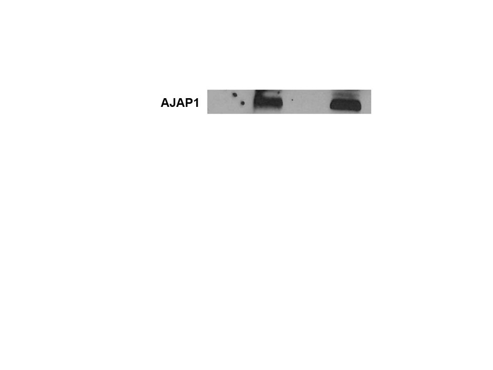
Detection of Mouse Shrew-1/AJAP1 by Western Blot. Western blot shows lysates of mouse brain (cortex) tissue, mouse brain (thalamus/hypothalamus) tissue, and mouse pancreas tissue. PVDF membrane was probed with 1 µg/mL of Sheep Anti-Mouse Shrew-1/AJAP1 Antigen Affinity-purified Polyclonal Antibody (Catalog # AF7970) followed by HRP-conjugated Anti-Sheep IgG Secondary Antibody (Catalog # HAF016). A specific band was detected for Shrew-1/ AJAP1 at approximately 45-50 kDa (as indicated). This experiment was conducted under reducing conditions and using Immunoblot Buffer Group 1.
Reconstitution Calculator
Preparation and Storage
- 12 months from date of receipt, -20 to -70 °C as supplied.
- 1 month, 2 to 8 °C under sterile conditions after reconstitution.
- 6 months, -20 to -70 °C under sterile conditions after reconstitution.
Background: Shrew-1/AJAP1
AJAP-1 (Adherens Junction Associated Protein 1; also Shrew-1 and Gm573) is a 44-47 kDa transmembrane protein found on the basolateral surface of epithelium. It is presumably expressed by multiple types of epithelium, and participates in cell-cell adhesion. Zonula adherens are structures that participate in the generation of cell contact adherens junctions. Such junctions, or complexes, are key to the maintenance of epithelial cell polarity and the maintenance of cell-to-cell contact. Central to this complex is E-cadherin, a transmembrane molecule that makes crucial contacts with cytoskeletal actin via catenins. AJAP-1 is an accompaning transmembrane molecule that interacts with E-cadherin and appears to promote the internalization of E-cadherin upon growth factor stimulation. This facilitates the dissolution of adherens junctions with an increase in cell motility. Notably, AJAP-1 is also known to complex with CD147 in nonpolar, invasive cells, suggesting that AJAP-1 may play a more complex role in cell migration. Mouse AJAP-1 is a 412 amino acid (aa) type III (no signal sequence) transmembrane protein. It contains a 284 aa extracellular region (aa 1-284) plus a 106 aa cytoplasmic domain (aa 306-412). Over aa 1-283, mouse AJAP-1 shares 95% and 75% aa sequence identity with rat and human AJAP-1, respectively; over aa 158-283, mouse AJAP-1 sequence identity changes little, showing 95% and 78% equality with rat and human AJAP-1, respectively.
Product Datasheets
Citations for Mouse Shrew-1/AJAP1 Antibody
R&D Systems personnel manually curate a database that contains references using R&D Systems products. The data collected includes not only links to publications in PubMed, but also provides information about sample types, species, and experimental conditions.
4
Citations: Showing 1 - 4
Filter your results:
Filter by:
-
Monoallelic de novo AJAP1 loss-of-function variants disrupt trans-synaptic control of neurotransmitter release
Authors: Früh, S;Boudkkazi, S;Koppensteiner, P;Sereikaite, V;Chen, LY;Fernandez-Fernandez, D;Rem, PD;Ulrich, D;Schwenk, J;Chen, Z;Le Monnier, E;Fritzius, T;Innocenti, SM;Besseyrias, V;Trovò, L;Stawarski, M;Argilli, E;Sherr, EH;van Bon, B;Kamsteeg, EJ;Iascone, M;Pilotta, A;Cutrì, MR;Azamian, MS;Hernández-García, A;Lalani, SR;Rosenfeld, JA;Zhao, X;Vogel, TP;Ona, H;Scott, DA;Scheiffele, P;Strømgaard, K;Tafti, M;Gassmann, M;Fakler, B;Shigemoto, R;Bettler, B;
Science advances
Species: Human, Mouse
Sample Types: Cell Lysates, Whole Tissue
Applications: Immunohistochemistry, Immunoprecipitation -
Complex formation of APP with GABAB receptors links axonal trafficking to amyloidogenic processing
Authors: MC Dinamarca, A Raveh, A Schneider, T Fritzius, S Früh, PD Rem, M Stawarski, T Lalanne, R Turecek, M Choo, V Besseyrias, W Bildl, D Bentrop, M Staufenbie, M Gassmann, B Fakler, J Schwenk, B Bettler
Nat Commun, 2019-03-22;10(1):1331.
Species: Human, Mouse
Sample Types: Transfected Whole Cells, Whole Tissue
Applications: Binding Assay, Immunoprecipitation, Western Blot -
Alternative exon usage creates novel transcript variants of tumor suppressor SHREW-1 gene with differential tissue expression profile
Authors: Anna Starzinski
Biol Open, 2016-11-15;5(11):1607-1619.
Species: Mouse
Sample Types: Tissue Homogenates, Whole Tissue
Applications: IHC-P, Immunoprecipitation -
Modular composition and dynamics of native GABAB receptors identified by high-resolution proteomics.
Authors: Schwenk J, Perez-Garci E, Schneider A, Kollewe A, Gauthier-Kemper A, Fritzius T, Raveh A, Dinamarca M, Hanuschkin A, Bildl W, Klingauf J, Gassmann M, Schulte U, Bettler B, Fakler B
Nat Neurosci, 2015-12-21;19(2):233-42.
Species: Mouse, Rat
Sample Types: Whole Cells
Applications: IHC-Fr
FAQs
No product specific FAQs exist for this product, however you may
View all Antibody FAQsReviews for Mouse Shrew-1/AJAP1 Antibody
Average Rating: 5 (Based on 2 Reviews)
Have you used Mouse Shrew-1/AJAP1 Antibody?
Submit a review and receive an Amazon gift card.
$25/€18/£15/$25CAN/¥75 Yuan/¥2500 Yen for a review with an image
$10/€7/£6/$10 CAD/¥70 Yuan/¥1110 Yen for a review without an image
Filter by:
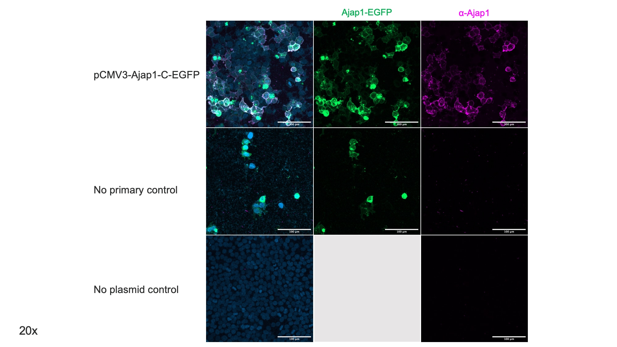
Cells were transfected with plasmids that express mouse Ajap1-C-EGFP. We diluted the Ajap1 antibody 1:500 in blocking buffer (10% donkey serum, 0.1% Tween 20 in PBS), and incubated overnight at 4ºC. We used a donkey anti-sheep-555 from abcam at 1:500 (same blocking buffer as above) with DAPI to stain nuclei.


