Mouse TGF-beta RIII Antibody Summary
Gly23-Thr785
Accession # NP_035708
Applications
Please Note: Optimal dilutions should be determined by each laboratory for each application. General Protocols are available in the Technical Information section on our website.
Scientific Data
 View Larger
Detection of Mouse TGF-beta RIII by Western Blot MicroRNA (miRNA) 466a-3p transfection inhibits regulatory T cell (Treg) polarization. Purified naïve CD4+ T cells were cultured under Treg-polarizing conditions along with the indicated mimic, control, or inhibitor conditions. Cells were harvested 48 h after addition of cytokines and miRNA mimics, inhibitors, or controls and subject to flow cytometry, immunoblot and quantitative real-time-PCR. The success of Treg polarization is examined as (A) representative dot plots gated on CD25HI cells and quantified in (B,C). Representative immunoblots of indicated proteins are presented in (D,F), along with associated densitometric measurements of transforming growth factor-beta 2 (TGF-beta 2) and TGF-beta R3 (E), and quantification of activated Smad 2, 3, and 4 (G). CD4+ cells were purified from naïve mouse lymph nodes and stimulated ex vivo with CD3 (3 µg/mL) and CD28 (3 µg/mL) for 48 h and administered Locked Nucleic Acid or controls at the time of seeding. Quantification of flow cytometry data from LAP-expressing FoxP3 positive Treg cells. (H) Purified naïve CD4+ T cells were cultured with either TGF-beta 1 (5 ng/mL) or TGF-beta 2 (5 ng/mL), along with CD3 (3 µg/mL), CD28 (3 µg/mL), and IL-2 (5 ng/mL) for 5 days. (I) representative dot plots of FoxP3, CD4-positive Tregs gated on CD25HI, (J), and their associated CD278 (ICOS) expression. Data are presented as mean ± SEM of three independent transfection experiments. *P < 0.05, **P < 0.005, ****P < 0.0001 by ANOVA with Tukey’s multiple comparison test. Image collected and cropped by CiteAb from the following publication (https://journal.frontiersin.org/article/10.3389/fimmu.2018.00688/full), licensed under a CC-BY license. Not internally tested by R&D Systems.
Reconstitution Calculator
Preparation and Storage
- 12 months from date of receipt, -20 to -70 °C as supplied.
- 1 month, 2 to 8 °C under sterile conditions after reconstitution.
- 6 months, -20 to -70 °C under sterile conditions after reconstitution.
Background: TGF-beta RIII
Transforming growth factor beta receptor III (TGF-beta RIII; also betaglycan) is a ubiquitously expressed, 280 kDa type I transmembrane proteoglycan member of the TGF-beta superfamily of proteins (1). Mouse TGF-beta RIII is synthesized as an 850 amino acid (aa) precursor that consists of a 22 aa signal sequence, a 763 extracellular domain (ECD), a 23 aa transmembrane region, and a 42 aa cytoplasmic tail. The large ECD contains heparan sulfate and chondroitin sulfate glycosaminoglycans, five potential N-linked glycosylation sites, and a zona pellucida-like domain from residues 454‑731 (1, 2). The short cytoplasmic domain is rich in serine and threonine, but has no discernible signaling structure typical of receptor kinases (2). Proteolysis at one of two potential juxtamembrane cleavage sites (Lys743Lys and Leu750AlaValVal) allows cells to release TGF-beta RIII in a soluble form (1, 2). Mouse TGF-beta RIII shares 94%, 82%, 80%, and 67% aa sequence identity with rat, human, porcine, and chicken TGF-beta RIII, respectively (2). In all of these species, TGF beta RIII contains 17 cysteines that are 100% conserved (2). TGF-beta RIII binds with high affinity to TGF-beta 1, TGF-beta 2, and TGF-beta 3 isoforms (1). TGF-beta RIII functions by binding, and then "presenting" ligand to TGF-beta type II receptors (1, 3). It also functions to limit ligand availability to the receptor via proteolysis which releases the soluble form of TGF beta RIII along with any bound factors, making them inaccessible to cell-surface receptors (1, 3). TGF-beta RIII can therefore enhance or inhibit cell signaling. TGF-beta RIII has been shown to play an essential role in the formation of the atrioventricular cushion and coronary vessels during development of the heart (4‑6). TGF beta RIII also plays a role in many cancers. Increased expression of TGF beta RIII is found in higher grade lymphomas, and reduced expression of TGF beta RIII is found with advanced stage neuroblastomas and ovarian carcinomas (4, 7‑9). Low TGF-beta RIII expression also correlates with higher grade among a cohort of breast cancers (4, 10). Additionally, overexpression of TGF-beta RIII in MDA-231 human breast cancer cells and DU145 prostate cancer cells results in decreased tumor invasion in vitro and in vivo (4, 11, 12).
- Kolodziejczyk, S.M. and B.K. Hall (1996) Biochem. Cell Biol. 74:299.
- Ponce-Castaneda, M.V. et al. (1998) Biochim. Biophys. Acta 1384:189.
- Lopez-Casillas, F. et al. (1993) Cell 73:1435.
- Criswell, T.L. and C.L. Arteaga (2007) J. Biol. Chem. 282:32491.
- Brown, C.B. et al. (1999) Science 283:2080.
- Compton, L.A. et al. (2007) Circ. Res. 101:784.
- Woszczyk, D. et al. (2004) Med. Sci. Monit. 10:CRIII3.
- Bristow, R.E. et al. (1999) Cancer 85:658.
- Iolascon, A. et al. (2000) Br. J. Cancer 82:1171.
- Dong, M. et al. (2007) J. Clin. Invest. 117:206.
- Turley, R.S. et al. (2007) Cancer Res. 67:1090.
- Sun, L. and C. Chen (1997) J. Biol. Chem. 272:25367.
Product Datasheets
Citations for Mouse TGF-beta RIII Antibody
R&D Systems personnel manually curate a database that contains references using R&D Systems products. The data collected includes not only links to publications in PubMed, but also provides information about sample types, species, and experimental conditions.
2
Citations: Showing 1 - 2
Filter your results:
Filter by:
-
miR-466a Targeting of TGF-beta 2 Contributes to FoxP3+ Regulatory T Cell Differentiation in a Murine Model of Allogeneic Transplantation
Authors: William Becker, Mitzi Nagarkatti, Prakash S. Nagarkatti
Frontiers in Immunology
-
Resveratrol protects mice against SEB-induced acute lung injury and mortality by miR-193a modulation that targets TGF-? signalling
Authors: H Alghetaa, A Mohammed, M Sultan, P Busbee, A Murphy, S Chatterjee, M Nagarkatti, P Nagarkatti
J. Cell. Mol. Med., 2018-03-07;0(0):.
Species: Mouse
Sample Types: Cell Lysates
Applications: Western Blot
FAQs
No product specific FAQs exist for this product, however you may
View all Antibody FAQsReviews for Mouse TGF-beta RIII Antibody
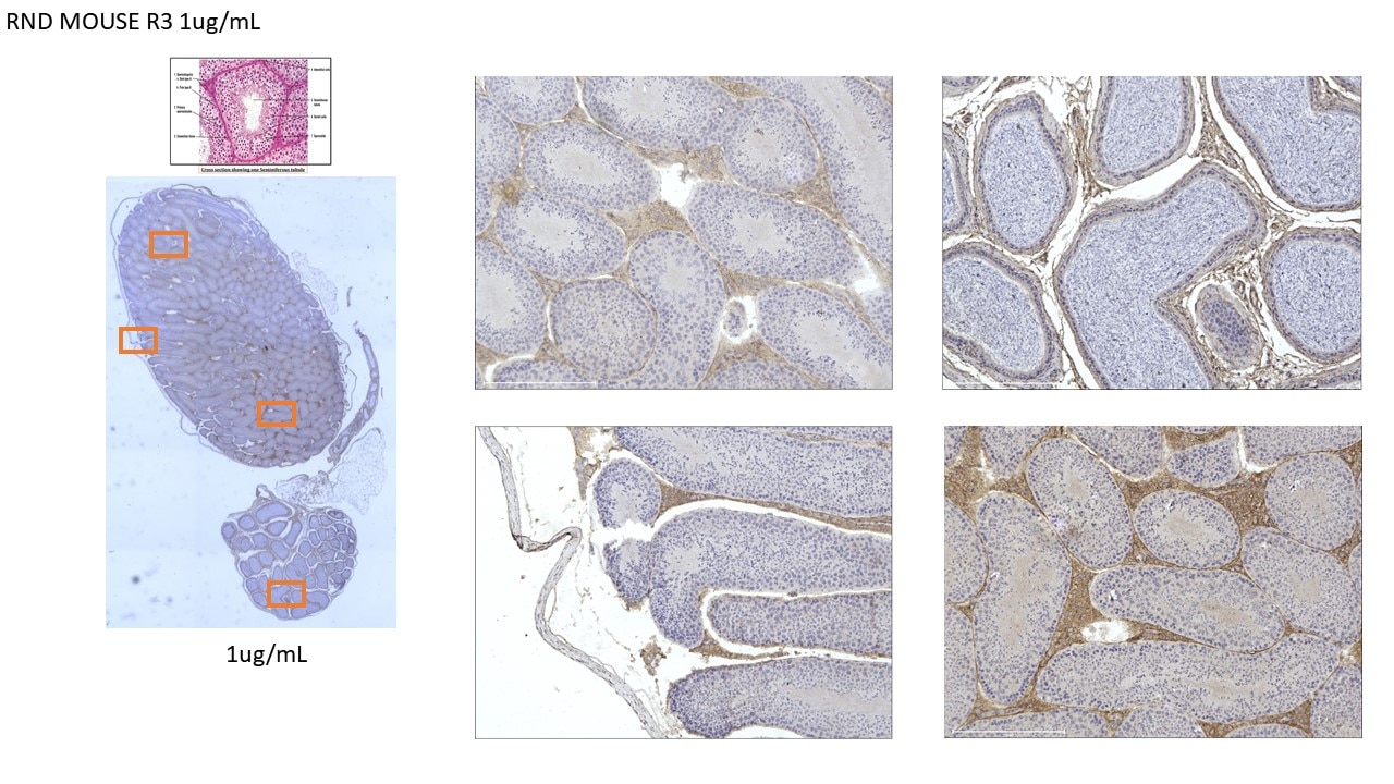
Average Rating: 4 (Based on 1 Review)
Have you used Mouse TGF-beta RIII Antibody?
Submit a review and receive an Amazon gift card.
$25/€18/£15/$25CAN/¥75 Yuan/¥2500 Yen for a review with an image
$10/€7/£6/$10 CAD/¥70 Yuan/¥1110 Yen for a review without an image
Filter by:


