SARS-CoV-2 Spike S2 Subunit Antibody
SARS-CoV-2 Spike S2 Subunit Antibody Summary
Met697-Pro1213
Applications
Please Note: Optimal dilutions should be determined by each laboratory for each application. General Protocols are available in the Technical Information section on our website.
Scientific Data
 View Larger
Detection of SARS-CoV-2 Spike S2 Subunit by Western Blot. Western blot shows recombinant SARS-CoV-2 Spike S2 protein. PVDF membrane was probed with 0.5 µg/mL of Rabbit Anti-SARS-CoV-2 Spike S2 Subunit Monoclonal Antibody (Catalog # MAB10850) followed by HRP-conjugated Anti-Rabbit IgG Secondary Antibody (HAF008). A specific band was detected for Spike S2 Subunit at approximately 75 kDa (as indicated). This experiment was conducted under reducing conditions and using Western Blot Buffer Group 1.
 View Larger
Spike S2 Subunit in HEK293 Human Cell Line Transfected with SARS-CoV-2. Spike S2 Subunit was detected in immersion fixed HEK293 human embryonic kidney cell line transfected with SARS-CoV-2 (positive staining) and HEK293 human embryonic kidney cell line (non-transfected, negative staining) using Rabbit Anti-SARS-CoV-2 Spike S2 Subunit Monoclonal Antibody (Catalog # MAB10850) at 3 µg/mL for 3 hours at room temperature. Cells were stained using the NorthernLights™ 557-conjugated Anti-Rabbit IgG Secondary Antibody (red; NL004) and counterstained with DAPI (blue). Specific staining was localized to cytoplasm. Staining was performed using our protocol for Fluorescent ICC Staining of Non-adherent Cells.
 View Larger
Spike S2 Subunit in SARS-CoV-2 infected Human lung. Spike S2 Subunit was detected in immersion fixed paraffin-embedded sections of SARS-CoV-2 infected human lung using Rabbit Anti-SARS-CoV-2 Spike S2 Subunit Monoclonal Antibody (Catalog # MAB10850) at 3 µg/mL for 1 hour at room temperature followed by incubation with the Anti-Rabbit IgG VisUCyte™ HRP Polymer Antibody (VC003). Before incubation with the primary antibody, tissue was subjected to heat-induced epitope retrieval using Antigen Retrieval Reagent-Basic (CTS013). Tissue was stained using DAB (brown) and counterstained with hematoxylin (blue). Specific staining was localized to immunoreactive profiles scattered throughout the tissue. Staining was performed using our protocol for IHC Staining with VisUCyte HRP Polymer Detection Reagents.
 View Larger
Detection of SARS-CoV-2 Spike Protein by Simple WesternTM. Simple Western lane view shows recombinant SARS-CoV-2 (positive sample) and OC43 coronavirus lysate (negative control), loaded at 0.2 mg/mL. A specific band was detected for the Spike Protein at approximately 268 kDa (as indicated) using 20 µg/mL of Rabbit Anti-SARS-CoV-2 Spike S2 Subunit Monoclonal Antibody (Catalog # MAB10850). This experiment was conducted under non-reducing conditions and using the 12-230 kDa separation system.
 View Larger
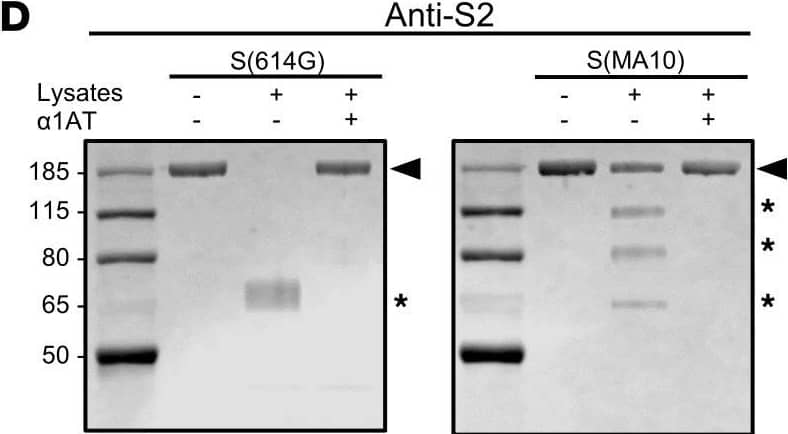
Detection of SARS-CoV-2 Spike S2 Subunit by Western Blot Neutrophil serine proteases degrade S and inhibit SARS-CoV-2 entry.(A and B) SDS-PAGE of recombinant trimeric S(614G) (A) and S(MA10) (B) incubated with purified human CatG, NE, and PR3 at indicated concentrations. Arrowheads indicate S protein and asterisks cleaved fragments. (C and D) Immunoblots of recombinant trimeric S(614G) and S(MA10) incubated with mouse neutrophil lysates with or without alpha 1AT and probed with anti-S1 (C) or anti-S2 (D) subunit antibodies. (E) Titers of VSV* delta G-S delta 21 incubated with NSPs. (F and G) Titers of SARS-CoV-2614G (F) and SARS-CoV-2MA10 (G) incubated with NSPs. Data in A–D representative of at least 3 independent experiments. Data in E–G were analyzed by 1-way ANOVA with Dunnett’s multiple-comparison test, comparing the NSP-treated group to the PBS control (n = 3). *P < 0.05, **P < 0.01, ***P < 0.001. Image collected and cropped by CiteAb from the following open publication (https://insight.jci.org/articles/view/174133), licensed under a CC-BY license. Not internally tested by R&D Systems.
Reconstitution Calculator
Preparation and Storage
- 12 months from date of receipt, -20 to -70 °C as supplied.
- 1 month, 2 to 8 °C under sterile conditions after reconstitution.
- 6 months, -20 to -70 °C under sterile conditions after reconstitution.
Background: Spike S2 Subunit
SARS-CoV-2, which causes the global pandemic coronavirus disease 2019 (Covid-19), belongs to a family of viruses known as coronaviruses that also include MERS and SARS-CoV-1. Coronaviruses are commonly comprised of four structural proteins: Spike protein(S), Envelope protein (E), Membrane protein (M) and Nucleocapsid protein (N) (1). SARS-CoV-2 Spike Protein (S Protein) is a glycoprotein that mediates membrane fusion and viral entry. The S protein is homotrimeric, with each ~180-kDa monomer consisting of two subunits, S1 and S2 (2). As with most coronaviruses, proteolytic cleavage of the S protein into two distinct peptides, S1 and S2 subunits, is required for activation. The S1 subunit is focused on attachment of the protein to the host receptor while the S2 subunit is involved with cell fusion (2-4). A metallopeptidase, angiotensin-converting enzyme 2 (ACE-2), has been identified as a functional receptor for SARS-CoV2, similar to SARS-CoV-1, through interaction with a receptor binding domain (RBD) located at the C-terminus of S1 subunit (5, 6). The S2 subunit of SARS-CoV-2 shares 90% and 41% amino acid sequence identity with the S2 subunit of SARS-CoV-1 and MERS, respectively. It has been demonstrated the S Protein can invade host cells through the CD147/EMMPRIN receptor and mediate membrane fusion (7, 8).
- Rota, P.A. et al. (2003) Science 300:1394.
- Bosch, B.J. et al. (2003). J. Virol. 77:8801.
- Belouzard, S. et al. (2009) Proc. Natl. Acad. Sci. USA 106:5871.
- Millet, J.K. and G. R. Whittaker (2015) Virus Res. 202:120.
- Li, W. et al. (2003) Nature 426:450.
- Wong, S.K. et al. (2004) J. Biol. Chem. 279:3197.
- Wang, X. et al. (2020) https://doi.org/10.1038/s41423-020-0424-9.
- Wang, K. et al. (2020) bioRxiv https://www.biorxiv.org/content/10.1101/2020.03.14.988345v.
Product Datasheets
FAQs
No product specific FAQs exist for this product, however you may
View all Antibody FAQsReviews for SARS-CoV-2 Spike S2 Subunit Antibody
There are currently no reviews for this product. Be the first to review SARS-CoV-2 Spike S2 Subunit Antibody and earn rewards!
Have you used SARS-CoV-2 Spike S2 Subunit Antibody?
Submit a review and receive an Amazon gift card.
$25/€18/£15/$25CAN/¥75 Yuan/¥2500 Yen for a review with an image
$10/€7/£6/$10 CAD/¥70 Yuan/¥1110 Yen for a review without an image
