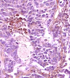
VEGF Receptor 1/Flt-1 in Human Breast Cancer Tissue.
Vascular Endothelial Growth Factor Receptor 1 (VEGF R1)/Flt-1 was detected in immersion fixed paraffin-embedded sections of human breast cancer tissue using Human VEGF R1 Antigen Affinity-purified Polyclonal Antibody (Catalog # AF321) at 15 µg/mL overnight at 4 °C. Tissue was stained (red) and counterstained with hematoxylin (blue). View our protocol for Chromogenic IHC Staining of Paraffin-embedded Tissue Sections.
This antibody specifically recognizes human VEGF R1. Reactivity with VEGF R1 from other species has not been determined.
Secondary Antibodies
R&D Systems offers a wide range of biotinylated, HRP-conjugated, fluorochrome-labeled, and unlabeled species-specific secondary antibodies. Our NorthernLights™ fluorescent secondary antibodies are available with three distinct excitation and emission maxima, making them ideal for multi-color fluorescence microscopy.
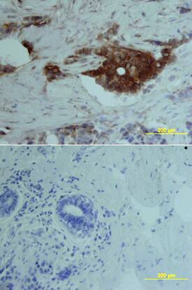
VEGF R1/Flt‑1 in Human Breast.
Vascular Endothelial Growth Factor Receptor 1 (VEGF R1/Flt‑1) was detected in immersion fixed paraffin-embedded sections of human breast array using Human VEGF R1/Flt‑1 Biotinylated Antigen Affinity-purified Polyclonal Antibody (Catalog # BAF321) at 15 µg/mL overnight at 4 °C. Tissue was stained using the Anti-Goat HRP-DAB Cell & Tissue Staining Kit (brown; Catalog # CTS008) and counterstained with hematoxylin (blue). Lower panel shows a lack of labeling if primary antibodies are omitted and tissue is stained only with secondary antibody followed by incubation with detection reagents. View our protocol for Chromogenic IHC Staining of Paraffin-embedded Tissue Sections.
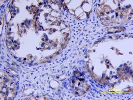
VEGF R1/Flt-1 in Human Ovarian Cancer Tissue.
Vascular Endothelial Growth Factor Receptor 1 (VEGF R1/Flt-1) was detected in immersion fixed paraffin-embedded sections of human ovarian cancer tissue using Human VEGF R1/Flt-1 Antigen Affinity-purified Polyclonal Antibody (Catalog # AF321) at 3 µg/mL overnight at 4 °C. Tissue was stained using the Anti-Goat HRP-DAB Cell & Tissue Staining Kit (brown; Catalog # CTS008) and counterstained with hematoxylin (blue). View our protocol for Chromogenic IHC Staining of Paraffin-embedded Tissue Sections.