J. Rivard, R. Weller Roska, N. Lee, A. James, K. Brumbaugh, and G. Wegner
ABSTRACT
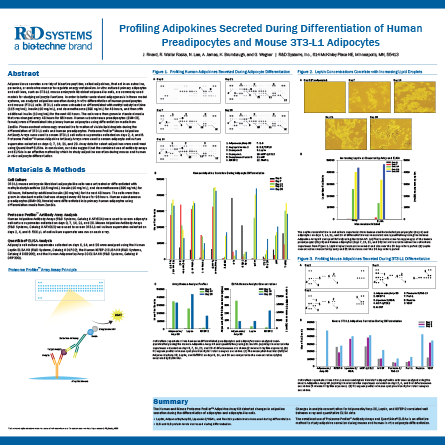
Adipose tissue secretes a variety of bioactive peptides, called adipokines, that act in an autocrine, paracrine, or endocrine manner to regulate energy metabolism. In vitro cultured primary adipocytes and cell lines, such as 3T3-L1 mouse embryonic fi broblast adipose-like cells, are commonly used models for studying adipocyte functions. In order to better understand adipogenesis in these model systems, we analyzed adipokine secretion during in vitro differentiation of human preadipocytes and mouse 3T3-L1 cells. 3T3-L1 cells were untreated or differentiated with methylisobutylxanthine (115 mg/mL), insulin (10 mg/mL), and dexamethasone (390 ng/mL) for 48 hours, and then with additional insulin (10 mg/mL) for the next 48 hours. The cells were then grown in standard media that was changed every 48 hours for 96 hours. Human subcutaneous preadipocytes (BMI=30, female) were differentiated into primary human adipocytes using differentiation media from ZenBio. Phase-contrast microscopy revealed the formation of visible lipid droplets during the differentiation of 3T3-L1 cells and human preadipocytes. Proteome Profiler™ Mouse Adipokine Antibody Arrays were used to screen 3T3-L1 cell culture supernates collected on days 2, 4, and 6. Proteome Profiler™ Human Adipokine Antibody Arrays were used to screen adipocyte cell culture supernates collected on days 0, 7, 14, 21, and 28. Array data for select adipokines were confirmed using Quantikine® ELISAs. In conclusion, our data suggest that the combined use of antibody arrays and ELISAs is an effective method by which to study adipokine secretion during mouse and human in vitro adipocyte differentiation.
