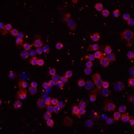
Human/Mouse/Rat Enolase 2/Neuron-specific Enolase Antibody
Human/Mouse/Rat Enolase 2/Neuron-specific Enolase Antibody Summary
Met1-Leu434
Accession # P09104
Customers also Viewed
Applications
Please Note: Optimal dilutions should be determined by each laboratory for each application. General Protocols are available in the Technical Information section on our website.
Scientific Data
 View Larger
Detection of Human/Mouse Enolase 2 by Western Blot. Western blot shows lysates of human brain, mouse brain tissue, and BG01V human embryonic stem cells. PVDF membrane was probed with 1 µg/mL of Human/Mouse Enolase 2 Antigen Affinity-purified Polyclonal Antibody (Catalog # AF5169) followed by HRP-conjugated Anti-Sheep IgG Secondary Antibody (Catalog # HAF016). A specific band was detected for Enolase 2 at approximately 46 kDa (as indicated). This experiment was conducted under reducing conditions and using Immunoblot Buffer Group 8.
 View Larger
Detection of Rat Enolase 2/Neuron‑specific Enolase by Western Blot. Western blot shows lysates of rat brain tissue, rat cerebellum tissue, and rat olfactroy bulb tissue. PVDF membrane was probed with 0.2 µg/mL of Sheep Anti-Human/Mouse Enolase 2/Neuron-specific Enolase Antigen Affinity-purified Polyclonal Antibody (Catalog # AF5169) followed by HRP-conjugated Anti-Sheep IgG Secondary Antibody (Catalog # HAF016). A specific band was detected for Enolase 2/Neuron-specific Enolase at approximately 47 kDa (as indicated). This experiment was conducted under reducing conditions and using Immunoblot Buffer Group 1.
 View Larger
Enolase 2 in Human Brain. Enolase 2 was detected in immersion fixed paraffin-embedded sections of human brain (cortex) using Human/Mouse Enolase 2 Antigen Affinity-purified Polyclonal Antibody (Catalog # AF5169) at 10 µg/mL overnight at 4 °C. Before incubation with the primary antibody, tissue was subjected to heat-induced epitope retrieval using Antigen Retrieval Reagent-Basic (Catalog # CTS013). Tissue was stained using the Anti-Sheep HRP-DAB Cell & Tissue Staining Kit (brown; Catalog # CTS019) and counterstained with hematoxylin (blue). Specific staining was localized to cytoplasm. View our protocol for Chromogenic IHC Staining of Paraffin-embedded Tissue Sections.
 View Larger
Detection of Human Enolase 2/Neuron‑specific Enolase by Simple WesternTM. Simple Western lane view shows lysates of BG01V human embryonic stem cells, loaded at 0.2 mg/mL. A specific band was detected for Enolase 2/Neuron-specific Enolase at approximately 50 kDa (as indicated) using 10 µg/mL of Sheep Anti-Human/Mouse Enolase 2/Neuron-specific Enolase Antigen Affinity-purified Polyclonal Antibody (Catalog # AF5169) followed by 1:50 dilution of HRP-conjugated Anti-Sheep IgG Secondary Antibody (Catalog # HAF016). This experiment was conducted under reducing conditions and using the 12-230 kDa separation system.
Preparation and Storage
- 12 months from date of receipt, -20 to -70 °C as supplied.
- 1 month, 2 to 8 °C under sterile conditions after reconstitution.
- 6 months, -20 to -70 °C under sterile conditions after reconstitution.
Background: Enolase 2/Neuron-specific Enolase
Enolase 2 (2-phospho-D-glycerate hydrolyase; also neural enolase and gamma -enolase) is a 46 kDa member of the Enolase family of enzymes. It is expressed in developing neurons and glia, is known to catalyze the generation of phosphoenolpyruvate, and is suggested to possess neurotrophic activity for neurons, likely through an extracellular mechanism. Human Enolase 2 is 434 amino acids (aa) in length. The enzymatic site spans most of the length of the molecule. Enolase 2 exists as both a noncovalently-linked homodimer, or heterodimer with alpha -enolase. Full-length human Enolase 2 is 99% aa identical to both mouse and canine Enolase 2. It shares 83% aa identity with human enolases # 1 and # 3.
Product Datasheets
Citations for Human/Mouse/Rat Enolase 2/Neuron-specific Enolase Antibody
R&D Systems personnel manually curate a database that contains references using R&D Systems products. The data collected includes not only links to publications in PubMed, but also provides information about sample types, species, and experimental conditions.
4
Citations: Showing 1 - 4
Filter your results:
Filter by:
-
Multivariate analysis of traumatic brain injury: development of an assessment score.
Authors: Buonora JE, Yarnell AM, Lazarus RC et al.
Front Neurol
-
VEGFA-modified DPSCs combined with LC-YE-PLGA NGCs promote facial nerve injury repair in rats
Authors: W Xu, X Xu, L Yao, B Xue, H Xi, X Cao, G Piao, S Lin, X Wang
Heliyon, 2023-03-28;9(4):e14626.
Species: Rat
Sample Types: Whole Tissue
Applications: IHC -
Methamphetamine leads to the alterations of microRNA profiles in the nucleus accumbens of rats
Authors: Jing Yang, Lihua Li, Shijun Hong, Dongxian Zhang, Yiqing Zhou
Pharmaceutical Biology
-
Inflammation-induced reversible switch of the neuron-specific enolase promoter from Purkinje neurons to Bergmann glia
Authors: Yusuke Sawada
Sci Rep, 2016-06-13;6(0):27758.
Species: Mouse
Sample Types: Whole Tissue
Applications: IHC-P
FAQs
No product specific FAQs exist for this product, however you may
View all Antibody FAQsIsotype Controls
Reconstitution Buffers
Secondary Antibodies
Reviews for Human/Mouse/Rat Enolase 2/Neuron-specific Enolase Antibody
Average Rating: 4 (Based on 4 Reviews)
Have you used Human/Mouse/Rat Enolase 2/Neuron-specific Enolase Antibody?
Submit a review and receive an Amazon gift card.
$25/€18/£15/$25CAN/¥75 Yuan/¥2500 Yen for a review with an image
$10/€7/£6/$10 CAD/¥70 Yuan/¥1110 Yen for a review without an image
Filter by:


