Human alpha-Smooth Muscle Actin APC-conjugated Antibody
Human alpha-Smooth Muscle Actin APC-conjugated Antibody Summary
Applications
Please Note: Optimal dilutions should be determined by each laboratory for each application. General Protocols are available in the Technical Information section on our website.
Scientific Data
 View Larger
Detection of alpha ‑Smooth Muscle Actin in Human Blood Monocytes by Flow Cytometry. Human peripheral blood monocytes were stained with Mouse Anti-Human a-Smooth Muscle Actin APC-conjugated Monoclonal Antibody (Catalog # IC1420A, filled histogram) or isotype control antibody (Catalog # IC003A, open histogram). To facilitate intracellular staining, cells were fixed with Flow Cytometry Fixation Buffer (Catalog # FC004) and permeabilized with Flow Cytometry Permeabilization/Wash Buffer I (Catalog # FC005). View our protocol for Staining Intracellular Molecules.
 View Larger
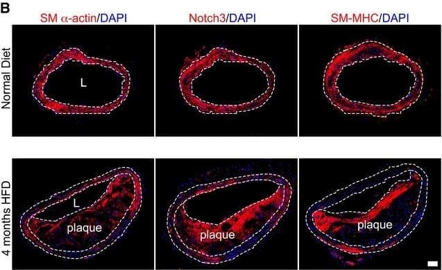
Detection of Mouse alpha-Smooth Muscle Actin by Immunohistochemistry FGFR1 and TGF beta signaling activity in smooth muscle cells in a mouse atherosclerosis modelADissected mouse aorta demonstrating lipid‐rich plaques in brachiocephalic artery after 4 months of high‐fat diet compared to the normal diet in Apoe−/− mice. Panels b, d are cross sections of brachiocephalic artery from a, c stained with Oil Red O. L: lumen. Scale bar: 4 mm. n = 3 mice per group.BHistological analysis of mouse normal artery or atherosclerotic plaque in brachiocephalic artery with anti‐SM alpha ‐actin, anti‐Notch3, and anti‐SM‐MHC antibodies. Nuclei were counterstained with DAPI (blue). L: lumen. Scale bar: 62 μm. n = 3 mice per group.C–FAnalysis of brachiocephalic artery of Apoe−/− mice maintained for 4 months on either normal or high‐fat diet using anti‐CD31 (green), anti‐p‐FGFR1 (red; C), anti‐FGFR1 (red; D), anti‐p‐Smad2 (red; E), and anti‐p‐Smad3 (red; F) antibodies. Nuclei were counterstained with DAPI (blue). Scale bar: 62 μm. L: lumen. M: media. n = 6 mice per group.G–JQuantification of the number of medial smooth muscle cells expressing p‐FGFR1, FGFR1, p‐Smad2, and p‐Smad3. ND: normal diet. HFD: high‐fat diet.Data information: All data shown as means ± SD (***P < 0.001 compared to ND, NS: not significant compared to ND; unpaired two‐tailed Student's t‐test). A full table of P‐values for this figure is shown in Appendix Table S1. Image collected and cropped by CiteAb from the following open publication (https://pubmed.ncbi.nlm.nih.gov/27189169), licensed under a CC-BY license. Not internally tested by R&D Systems.
Reconstitution Calculator
Preparation and Storage
- 12 months from date of receipt, 2 to 8 °C as supplied.
Background: alpha-Smooth Muscle Actin
alpha ‑Smooth Muscle Actin has been frequently used as a marker of smooth muscle differentiation (1, 2).
- Skalli, O. et al. (1986) J. Cell Biol. 103:2787.
- Oishi, K. et al. (2002) J. Physiol. 540:139.
Product Datasheets
Citations for Human alpha-Smooth Muscle Actin APC-conjugated Antibody
R&D Systems personnel manually curate a database that contains references using R&D Systems products. The data collected includes not only links to publications in PubMed, but also provides information about sample types, species, and experimental conditions.
15
Citations: Showing 1 - 10
Filter your results:
Filter by:
-
A human multi-cellular model shows how platelets drive production of diseased extracellular matrix and tissue invasion
Authors: Malacrida B, Nichols S, Maniati E et al.
iScience
-
Inhibition of the mTOR pathway in abdominal aortic aneurysm: implications of smooth muscle cell contractile phenotype, inflammation, and aneurysm expansion
Authors: Guangxin Li, Lingfeng Qin, Lei Wang, Xuan Li, Alexander W. Caulk, Jian Zhang et al.
American Journal of Physiology-Heart and Circulatory Physiology
-
Modelling TGF beta R and Hh pathway regulation of prognostic matrisome molecules in ovarian cancer
Authors: Delaine-Smith R, Maniati E, Malacrida B et al.
iScience
-
Mechanostimulatory cues determine intestinal fibroblast fate and profibrotic remodeling in a physiodynamic human gut-on-a-chip
Authors: Min, S;Than, N;Shin, YC;Ertugral, EG;Kothapalli, CR;Awoniyi, O;Kim, HJ;
bioRxiv : the preprint server for biology
Species: Human
Sample Types: Whole Cells
Applications: Flow Cytometry -
Mesothelial cells with mesenchymal features enhance peritoneal dissemination by forming a protumorigenic microenvironment
Authors: Yonemura, A;Semba, T;Zhang, J;Fan, Y;Yasuda-Yoshihara, N;Wang, H;Uchihara, T;Yasuda, T;Nishimura, A;Fu, L;Hu, X;Wei, F;Kitamura, F;Akiyama, T;Yamashita, K;Eto, K;Iwagami, S;Iwatsuki, M;Miyamoto, Y;Matsusaki, K;Yamasaki, J;Nagano, O;Saya, H;Song, S;Tan, P;Baba, H;Ajani, JA;Ishimoto, T;
Cell reports
Species: Mouse
Sample Types: Whole Cells
Applications: Flow Cytometry -
Orthotopic model of pancreatic cancer using CD34+ humanized mice and generation of tumor organoids from humanized tumors
Authors: Hye Jeong, J;Park, S;Lee, S;Kim, Y;Kyong Shim, I;Jeong, SY;Kyung Choi, E;Kim, J;Jun, E;
International immunopharmacology
Species: Mouse
Sample Types: Whole Cells
Applications: Flow Cytometry -
Targeting Periostin Expression Makes Pancreatic Cancer Spheroids More Vulnerable to Natural Killer Cells
Authors: D Karakas, M Erkisa, RO Akar, G Akman, EY Senol, E Ulukaya
Biomedicines, 2023-01-19;11(2):.
Species: Human
Sample Types: Whole Cells
Applications: Flow Cytometry -
microRNA expression profile in Smooth Muscle Cells isolated from thoracic aortic aneurysm samples
Authors: A Kasprzyk-P, A Wojciechow, M Kuc, J Zielinski, A Parulski, M Kusmierczy, A Lutynska, K Kozar-Kami
Adv Med Sci, 2019-04-22;64(2):331-337.
Species: Human
Sample Types: Whole Cells
Applications: Flow Cytometry -
Adipose-tissue-derived therapeutic cells in their natural environment as an autologous cell therapy strategy: the microtissue-stromal vascular fraction
Authors: S Nürnberger, C Lindner, J Maier, K Strohmeier, C Wurzer, P Slezak, S Suessner, W Holnthoner, H Redl, S Wolbank, E Priglinger
Eur Cell Mater, 2019-02-22;37(0):113-133.
Species: Human
Sample Types: Whole Cells
Applications: Flow Cytometry -
Transcriptome network analysis identifies protective role of the LXR/SREBP-1c axis in murine pulmonary fibrosis
Authors: S Shichino, S Ueha, S Hashimoto, M Otsuji, J Abe, T Tsukui, S Deshimaru, T Nakajima, M Kosugi-Kan, FH Shand, Y Inagaki, H Shimano, K Matsushima
JCI Insight, 2019-01-10;4(1):.
Species: Mouse
Sample Types: Tissue Homogenates
Applications: Flow Cytometry -
Lung fibroblasts express a miR-19a-19b-20a sub-cluster to suppress TGF-?-associated fibroblast activation in murine pulmonary fibrosis
Authors: K Souma, S Shichino, S Hashimoto, S Ueha, T Tsukui, T Nakajima, HI Suzuki, FHW Shand, Y Inagaki, T Nagase, K Matsushima
Sci Rep, 2018-11-09;8(1):16642.
Species: Mouse
Sample Types: Whole Cells
Applications: Flow Cytometry -
Mutational analysis of AKT1 and PIK3CA in intraductal papillomas of the breast with special reference to cellular components
Authors: C Mishima, N Kagara, JI Ikeda, E Morii, T Miyake, T Tanei, Y Naoi, M Shimoda, K Shimazu, SJ Kim, S Noguchi
Am. J. Pathol., 2018-02-16;0(0):.
Species: Human
Sample Types: Whole Cells
Applications: Flow Cytometry -
Fibroblast Heterogeneity and Immunosuppressive Environment in Human Breast Cancer
Authors: A Costa, Y Kieffer, A Scholer-Da, F Pelon, B Bourachot, M Cardon, P Sirven, I Magagna, L Fuhrmann, C Bernard, C Bonneau, M Kondratova, I Kuperstein, A Zinovyev, AM Givel, MC Parrini, V Soumelis, A Vincent-Sa, F Mechta-Gri
Cancer Cell, 2018-02-15;0(0):.
Species: Human
Sample Types: Whole Cells
Applications: FACS -
High-throughput immunophenotypic characterization of bone marrow- and cord blood-derived mesenchymal stromal cells reveals common and differentially expressed markers: identification of angiotensin-converting enzyme (CD143) as a marker differentially expressed between adult and perinatal tissue sources
Authors: E Amati, O Perbellini, G Rotta, M Bernardi, K Chieregato, S Sella, F Rodeghiero, M Ruggeri, G Astori
Stem Cell Res Ther, 2018-01-16;9(1):10.
Species: Human
Sample Types: Whole Cells
Applications: Flow Cytometry -
Endothelial-to-mesenchymal transition drives atherosclerosis progression.
Authors: Chen PY, Qin L, Baeyens N et al.
J Clin Invest
FAQs
No product specific FAQs exist for this product, however you may
View all Antibody FAQsReviews for Human alpha-Smooth Muscle Actin APC-conjugated Antibody
There are currently no reviews for this product. Be the first to review Human alpha-Smooth Muscle Actin APC-conjugated Antibody and earn rewards!
Have you used Human alpha-Smooth Muscle Actin APC-conjugated Antibody?
Submit a review and receive an Amazon gift card.
$25/€18/£15/$25CAN/¥75 Yuan/¥2500 Yen for a review with an image
$10/€7/£6/$10 CAD/¥70 Yuan/¥1110 Yen for a review without an image

