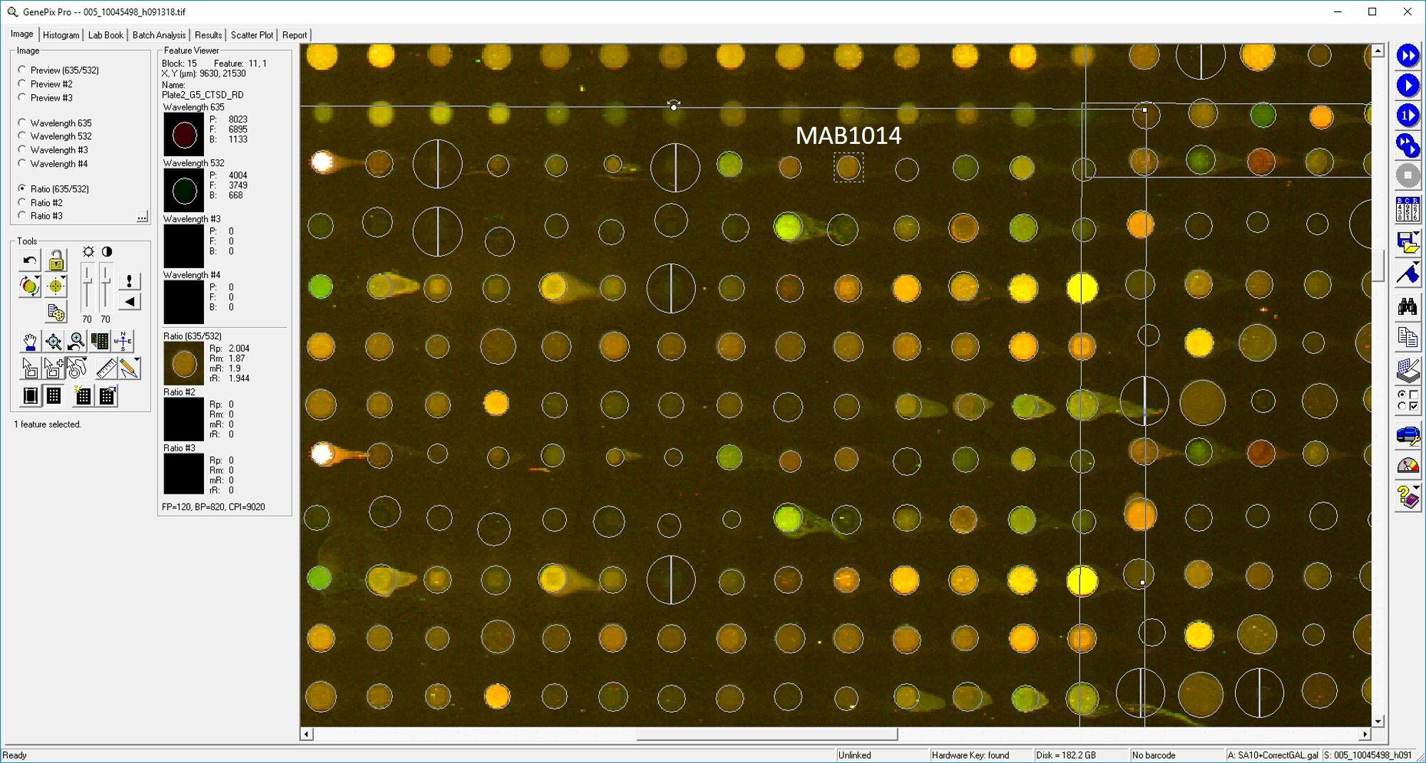
Human Cathepsin D Antibody Summary
Leu21-Leu412
Accession # P07339
Applications
Please Note: Optimal dilutions should be determined by each laboratory for each application. General Protocols are available in the Technical Information section on our website.
Scientific Data
 View Larger
Detection of Human Cathepsin D by Western Blot. Western blot shows lysates of PANC-1 human pancreatic carcinoma cell line and MCF-7 human breast cancer cell line. PVDF membrane was probed with 0.2 µg/mL of Mouse Anti-Human Cathepsin D Monoclonal Antibody (Catalog # MAB1014) followed by HRP-conjugated Anti-Mouse IgG Secondary Antibody (Catalog # HAF018). Specific bands were detected for Cathepsin D at approximately 28 and 46 kDa (as indicated). This experiment was conducted under reducing conditions and using Immunoblot Buffer Group 1.
 View Larger
Cathepsin D in Human Prostate Cancer Tissue. Cathepsin D was detected in immersion fixed paraffin-embedded sections of human prostate cancer tissue using Mouse Anti-Human Cathepsin D Monoclonal Antibody (Catalog # MAB1014) at 5 µg/mL for 1 hour at room temperature followed by incubation with the Anti-Mouse IgG VisUCyte™ HRP Polymer Antibody (Catalog # VC001). Tissue was stained using DAB (brown) and counterstained with hematoxylin (blue). Specific staining was localized to epithelial cell cytoplasm. View our protocol for IHC Staining with VisUCyte HRP Polymer Detection Reagents.
 View Larger
Detection of Human Cathepsin D by Simple WesternTM. Simple Western lane view shows lysates of PANC‑1 human pancreatic carcinoma cell line and MCF‑7 human breast cancer cell line, loaded at 0.2 mg/mL. Specific bands were detected for Cathepsin D at approximately 36 and 52-57 kDa (as indicated) using 2 µg/mL of Mouse Anti-Human Cathepsin D Monoclonal Antibody (Catalog # MAB1014). This experiment was conducted under reducing conditions and using the 12-230 kDa separation system.
Reconstitution Calculator
Preparation and Storage
- 12 months from date of receipt, -20 to -70 °C as supplied.
- 1 month, 2 to 8 °C under sterile conditions after reconstitution.
- 6 months, -20 to -70 °C under sterile conditions after reconstitution.
Background: Cathepsin D
Cathepsin D is a lysosomal aspartic protease of the pepsin family (1). Human cathepsin D is synthesized as a precursor protein, consisting of a signal peptide (aa 1‑18), a propeptide (aa 19-64), and a mature chain (aa 65‑412) (2‑4). The mature chain can be processed further to the light (aa 65‑161) and heavy (aa 169‑412) chains. It is expressed in most cells and overexpressed in breast cancer cells (5). It is a major enzyme in protein degradation in lysosomes, and also involved in the presentation of antigenic peptides. Mice deficient in this enzyme showed a progressive atrophy of the intestinal mucosa, a massive destruction of lymphoid organs, and a profound neuronal ceroid lipofucinosis, indicating that cathepsin D is essential for proteolysis of proteins regulating cell growth and tissue homeostasis (6). Cathepsin D secreted from human prostate carcinoma cells are responsible for the generation of angiostatin, a potent endogeneous inhibitor of angiogenesis (6).
- Conner et al. in Handbook of Proteolytic Enzymes Barrett (1998) Academic Press, San Diego, p. 828.
- Faust, et al. (1985) Proc. Natl. Acad. Sci. USA 82:4910.
- Westley and May (1987) Nucl. Acid Res. 15:3773.
- Redecker, et al. (1991) DNA Cell Biol. 10:423.
- Rochefort, et al. (2000) Clin. Chim. Acta. 291:157.
- Tsukuba, et al. (2000) Mol. Cells 10:601.
Product Datasheets
Citation for Human Cathepsin D Antibody
R&D Systems personnel manually curate a database that contains references using R&D Systems products. The data collected includes not only links to publications in PubMed, but also provides information about sample types, species, and experimental conditions.
1 Citation: Showing 1 - 1
-
Biomarker discovery from pancreatic cancer secretome using a differential proteomic approach.
Authors: Gronborg M, Kristiansen TZ, Iwahori A, Chang R, Reddy R, Sato N, Molina H, Jensen ON, Hruban RH, Goggins MG, Maitra A, Pandey A
Mol. Cell Proteomics, 2005-10-08;5(1):157-71.
Species: Human
Sample Types: Cell Lysates
Applications: Western Blot
FAQs
No product specific FAQs exist for this product, however you may
View all Antibody FAQsReviews for Human Cathepsin D Antibody
Average Rating: 3.8 (Based on 4 Reviews)
Have you used Human Cathepsin D Antibody?
Submit a review and receive an Amazon gift card.
$25/€18/£15/$25CAN/¥75 Yuan/¥2500 Yen for a review with an image
$10/€7/£6/$10 CAD/¥70 Yuan/¥1110 Yen for a review without an image
Filter by:


