Human CD34 APC-conjugated Antibody Summary
Applications
Please Note: Optimal dilutions should be determined by each laboratory for each application. General Protocols are available in the Technical Information section on our website.
Scientific Data
 View Larger
Detection of CD34 in Human PBMCs by Flow Cytometry. Human peripheral blood mononuclear cells (PBMCs) were stained with Mouse Anti-Human CD34 APC-conjugated Monoclonal Antibody (Catalog # FAB7227A) and Mouse Anti-Human CD45 PE-conjugated Monoclonal Antibody (Catalog # FAB1430P). Quadrant markers were set based on control antibody staining (Catalog # IC002A). View our protocol for Staining Membrane-associated Proteins.
 View Larger
Detection of CD34 in KG‑1a Human Cell Line by Flow Cytometry. KG-1a human acute myelogenous leukemia cell line was stained with Mouse Anti-Human CD34 APC-conjugated Monoclonal Antibody (Catalog # FAB7227A, filled histogram) or isotype control antibody (Catalog # IC002A, open histogram). View our protocol for Staining Membrane-associated Proteins.
 View Larger
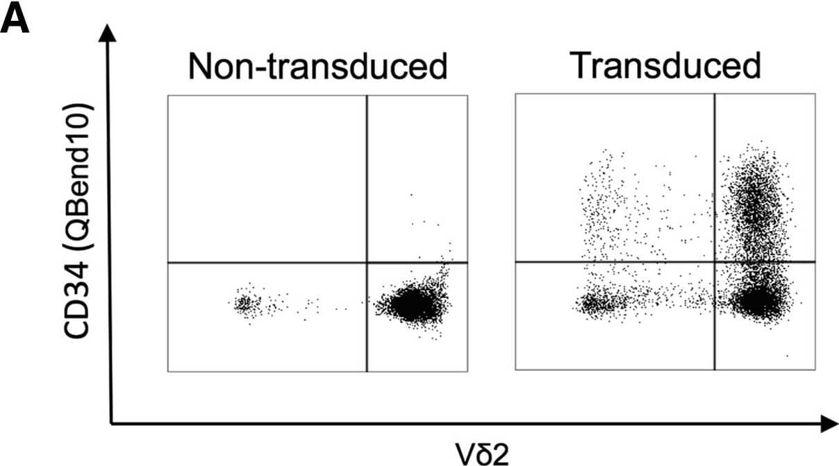
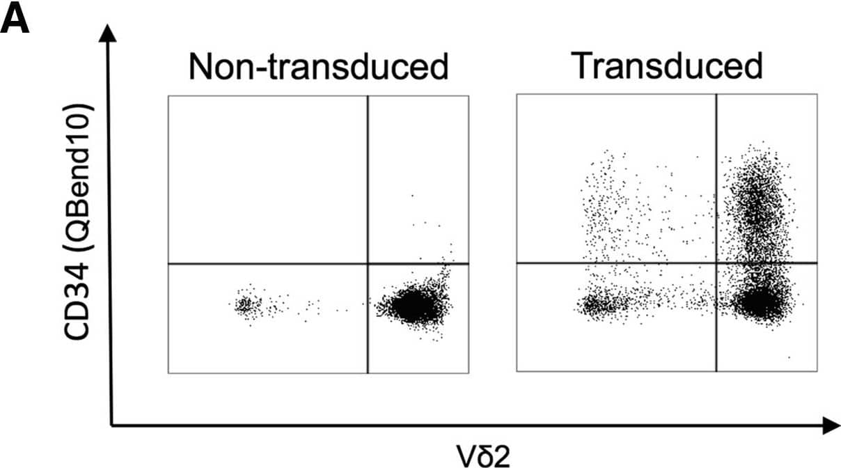
Detection of Human CD34 by Flow Cytometry alpha beta, Vδ1+, and Vδ2+ T Cells Are Efficiently Transduced with GD2-CAR following Activation with CD3/CD28 Antibody, ZOL, or ConA, and Bulk Populations Are Cytotoxic to Neuroblastoma Cells(A) Representative flow cytometry dot plot showing transduction efficiency of ZOL-expanded non-transduced and GD2-CAR+-transduced PBMCs. Vδ2+ populations were gated on CD3+ live cells 8 days following transduction. The GD2-CAR construct coexpresses the QBend10 epitope from CD34, allowing detection by flow cytometry. Transduction efficiency was determined by the percentage of QBend10+ in the T cell population gate compared to control non-transduced cells. (B–D) Mean transduction efficiency using CD3/CD28 antibody, ZOL, or ConA activation methods, respectively. Each data point represents an individual donor (n = 9) and each horizontal line is the mean. (E–G) Bulk populations of GD2-CAR-transduced T cells stimulated by CD3/CD28 antibody (E), ZOL (F), and ConA (G) specifically lyse the GD2-expressing neuroblastoma cell line, LAN1, in 4-hr 51Cr release assay. NTD, non-transduced T cells; TD, transduced GD2-CAR+ T cells (data represented as mean ± SEM; 3–5 individual donors in triplicate). Image collected and cropped by CiteAb from the following publication (https://linkinghub.elsevier.com/retrieve/pii/S1525001617305981), licensed under a CC-BY license. Not internally tested by R&D Systems.
 View Larger
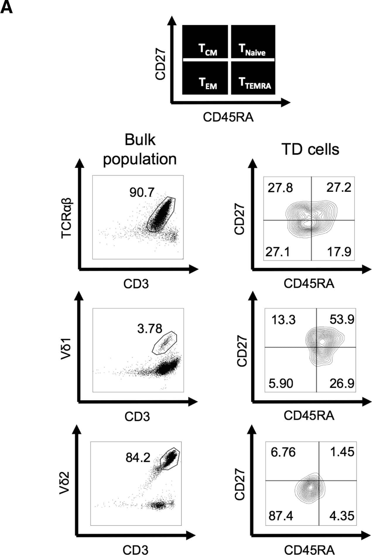
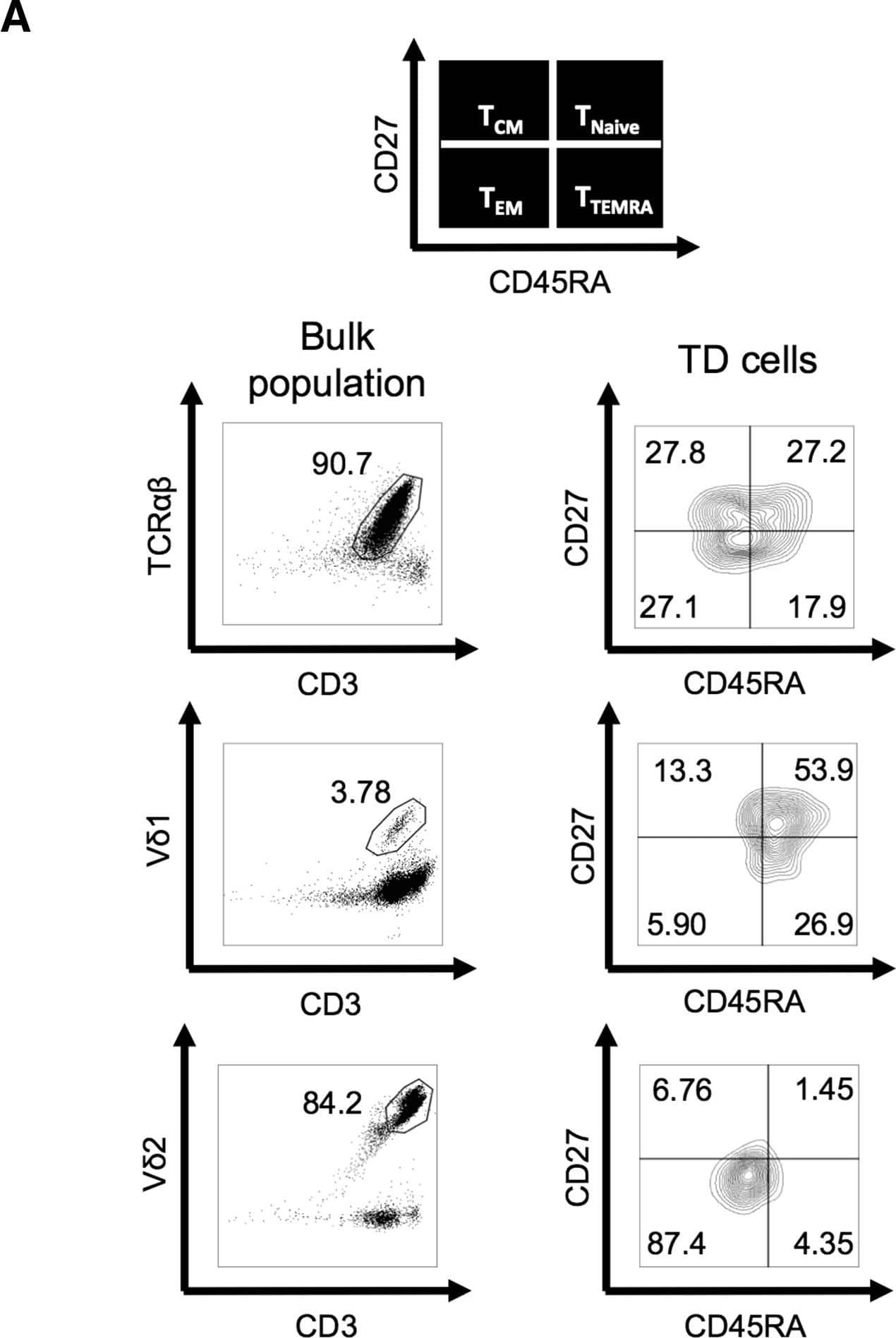
Detection of Human CD34 by Flow Cytometry Memory Phenotype and Exhaustion Marker Expression on Expanded CD3+ alpha beta, Vδ1+, and Vδ2+ Live T Cells(A) Representative flow cytometry dot plot showing gating of T cell subtypes ( alpha beta, Vδ1+, and Vδ2+) and contour plots of the corresponding subtype memory phenotype (N, naive; CM, central memory; EM, effector memory; TEMRA, terminally differentiated effector memory) according to expression of CD27 and CD45RA. (B–D) memory phenotypes of pre-expanded PBMCs (B), post-expansion non-transduced (NTD) cells (C), and post-expansion GD2-CAR-transduced cells (D). alpha beta cells were stimulated with CD3/CD28 antibody, Vδ1 cells were stimulated with ConA, and Vδ2 cells were stimulated with ZOL (data represented as mean ± SEM; alpha beta and Vδ2, n = 6; Vδ1, n = 3). (E and F) Expanded Vδ1+ cells transduced with CAR after ConA stimulation express significantly fewer exhaustion markers (PD1 and TIM3) than alpha beta and Vδ2+ CAR-T cells. (E) Flow cytometry plots from a representative donor. alpha beta cells were stimulated with CD3/CD28 antibody, Vδ1+ cells were stimulated with ConA, and Vδ2+ cells were stimulated with ZOL. (F) PD1 and TIM3 expression on TD and NTD T cells 13 days following activation (data represented as mean ± SEM; alpha beta and Vδ2+, n = 6; Vδ1+, n = 3; p value for significance comparing double-negative populations). Image collected and cropped by CiteAb from the following publication (https://linkinghub.elsevier.com/retrieve/pii/S1525001617305981), licensed under a CC-BY license. Not internally tested by R&D Systems.
 View Larger
Detection of Human Human CD34 APC-conjugated Antibody by Flow Cytometry alpha beta, Vδ1+, and Vδ2+ T Cells Are Efficiently Transduced with GD2-CAR following Activation with CD3/CD28 Antibody, ZOL, or ConA, and Bulk Populations Are Cytotoxic to Neuroblastoma Cells(A) Representative flow cytometry dot plot showing transduction efficiency of ZOL-expanded non-transduced and GD2-CAR+-transduced PBMCs. Vδ2+ populations were gated on CD3+ live cells 8 days following transduction. The GD2-CAR construct coexpresses the QBend10 epitope from CD34, allowing detection by flow cytometry. Transduction efficiency was determined by the percentage of QBend10+ in the T cell population gate compared to control non-transduced cells. (B–D) Mean transduction efficiency using CD3/CD28 antibody, ZOL, or ConA activation methods, respectively. Each data point represents an individual donor (n = 9) and each horizontal line is the mean. (E–G) Bulk populations of GD2-CAR-transduced T cells stimulated by CD3/CD28 antibody (E), ZOL (F), and ConA (G) specifically lyse the GD2-expressing neuroblastoma cell line, LAN1, in 4-hr 51Cr release assay. NTD, non-transduced T cells; TD, transduced GD2-CAR+ T cells (data represented as mean ± SEM; 3–5 individual donors in triplicate). Image collected and cropped by CiteAb from the following publication (https://pubmed.ncbi.nlm.nih.gov/29310916), licensed under a CC-BY license. Not internally tested by R&D Systems.
 View Larger
Detection of Human Human CD34 APC-conjugated Antibody by Flow Cytometry Memory Phenotype and Exhaustion Marker Expression on Expanded CD3+ alpha beta, Vδ1+, and Vδ2+ Live T Cells(A) Representative flow cytometry dot plot showing gating of T cell subtypes ( alpha beta, Vδ1+, and Vδ2+) and contour plots of the corresponding subtype memory phenotype (N, naive; CM, central memory; EM, effector memory; TEMRA, terminally differentiated effector memory) according to expression of CD27 and CD45RA. (B–D) memory phenotypes of pre-expanded PBMCs (B), post-expansion non-transduced (NTD) cells (C), and post-expansion GD2-CAR-transduced cells (D). alpha beta cells were stimulated with CD3/CD28 antibody, Vδ1 cells were stimulated with ConA, and Vδ2 cells were stimulated with ZOL (data represented as mean ± SEM; alpha beta and Vδ2, n = 6; Vδ1, n = 3). (E and F) Expanded Vδ1+ cells transduced with CAR after ConA stimulation express significantly fewer exhaustion markers (PD1 and TIM3) than alpha beta and Vδ2+ CAR-T cells. (E) Flow cytometry plots from a representative donor. alpha beta cells were stimulated with CD3/CD28 antibody, Vδ1+ cells were stimulated with ConA, and Vδ2+ cells were stimulated with ZOL. (F) PD1 and TIM3 expression on TD and NTD T cells 13 days following activation (data represented as mean ± SEM; alpha beta and Vδ2+, n = 6; Vδ1+, n = 3; p value for significance comparing double-negative populations). Image collected and cropped by CiteAb from the following publication (https://pubmed.ncbi.nlm.nih.gov/29310916), licensed under a CC-BY license. Not internally tested by R&D Systems.
 View Larger
Detection of Human CD34 by Flow Cytometry alpha beta, Vδ1+, and Vδ2+ T Cells Are Efficiently Transduced with GD2-CAR following Activation with CD3/CD28 Antibody, ZOL, or ConA, and Bulk Populations Are Cytotoxic to Neuroblastoma Cells(A) Representative flow cytometry dot plot showing transduction efficiency of ZOL-expanded non-transduced and GD2-CAR+-transduced PBMCs. Vδ2+ populations were gated on CD3+ live cells 8 days following transduction. The GD2-CAR construct coexpresses the QBend10 epitope from CD34, allowing detection by flow cytometry. Transduction efficiency was determined by the percentage of QBend10+ in the T cell population gate compared to control non-transduced cells. (B–D) Mean transduction efficiency using CD3/CD28 antibody, ZOL, or ConA activation methods, respectively. Each data point represents an individual donor (n = 9) and each horizontal line is the mean. (E–G) Bulk populations of GD2-CAR-transduced T cells stimulated by CD3/CD28 antibody (E), ZOL (F), and ConA (G) specifically lyse the GD2-expressing neuroblastoma cell line, LAN1, in 4-hr 51Cr release assay. NTD, non-transduced T cells; TD, transduced GD2-CAR+ T cells (data represented as mean ± SEM; 3–5 individual donors in triplicate). Image collected and cropped by CiteAb from the following open publication (https://pubmed.ncbi.nlm.nih.gov/29310916), licensed under a CC-BY license. Not internally tested by R&D Systems.
 View Larger
Detection of Human CD34 by Flow Cytometry Memory Phenotype and Exhaustion Marker Expression on Expanded CD3+ alpha beta, Vδ1+, and Vδ2+ Live T Cells(A) Representative flow cytometry dot plot showing gating of T cell subtypes ( alpha beta, Vδ1+, and Vδ2+) and contour plots of the corresponding subtype memory phenotype (N, naive; CM, central memory; EM, effector memory; TEMRA, terminally differentiated effector memory) according to expression of CD27 and CD45RA. (B–D) memory phenotypes of pre-expanded PBMCs (B), post-expansion non-transduced (NTD) cells (C), and post-expansion GD2-CAR-transduced cells (D). alpha beta cells were stimulated with CD3/CD28 antibody, Vδ1 cells were stimulated with ConA, and Vδ2 cells were stimulated with ZOL (data represented as mean ± SEM; alpha beta and Vδ2, n = 6; Vδ1, n = 3). (E and F) Expanded Vδ1+ cells transduced with CAR after ConA stimulation express significantly fewer exhaustion markers (PD1 and TIM3) than alpha beta and Vδ2+ CAR-T cells. (E) Flow cytometry plots from a representative donor. alpha beta cells were stimulated with CD3/CD28 antibody, Vδ1+ cells were stimulated with ConA, and Vδ2+ cells were stimulated with ZOL. (F) PD1 and TIM3 expression on TD and NTD T cells 13 days following activation (data represented as mean ± SEM; alpha beta and Vδ2+, n = 6; Vδ1+, n = 3; p value for significance comparing double-negative populations). Image collected and cropped by CiteAb from the following open publication (https://pubmed.ncbi.nlm.nih.gov/29310916), licensed under a CC-BY license. Not internally tested by R&D Systems.
 View Larger
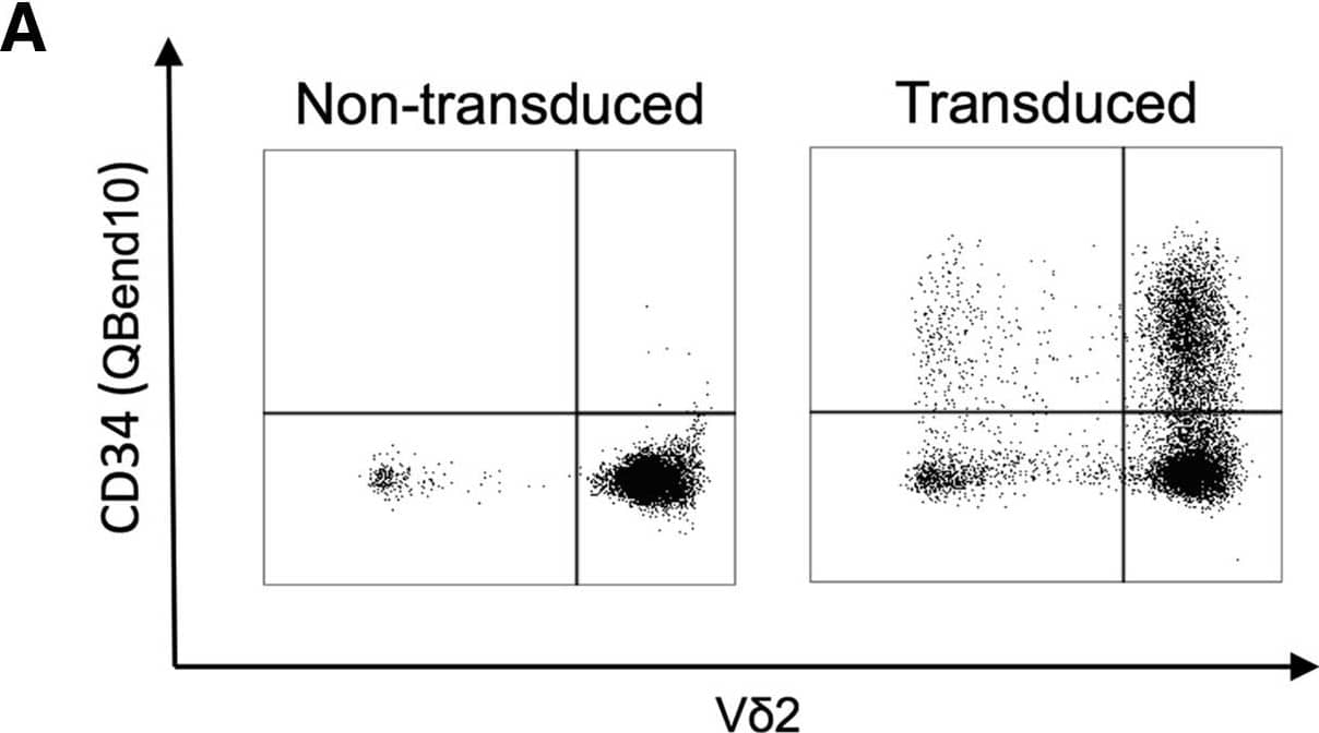
Detection of Human CD34 by Flow Cytometry alpha beta, Vδ1+, and Vδ2+ T Cells Are Efficiently Transduced with GD2-CAR following Activation with CD3/CD28 Antibody, ZOL, or ConA, and Bulk Populations Are Cytotoxic to Neuroblastoma Cells(A) Representative flow cytometry dot plot showing transduction efficiency of ZOL-expanded non-transduced and GD2-CAR+-transduced PBMCs. Vδ2+ populations were gated on CD3+ live cells 8 days following transduction. The GD2-CAR construct coexpresses the QBend10 epitope from CD34, allowing detection by flow cytometry. Transduction efficiency was determined by the percentage of QBend10+ in the T cell population gate compared to control non-transduced cells. (B–D) Mean transduction efficiency using CD3/CD28 antibody, ZOL, or ConA activation methods, respectively. Each data point represents an individual donor (n = 9) and each horizontal line is the mean. (E–G) Bulk populations of GD2-CAR-transduced T cells stimulated by CD3/CD28 antibody (E), ZOL (F), and ConA (G) specifically lyse the GD2-expressing neuroblastoma cell line, LAN1, in 4-hr 51Cr release assay. NTD, non-transduced T cells; TD, transduced GD2-CAR+ T cells (data represented as mean ± SEM; 3–5 individual donors in triplicate). Image collected and cropped by CiteAb from the following open publication (https://pubmed.ncbi.nlm.nih.gov/29310916), licensed under a CC-BY license. Not internally tested by R&D Systems.
 View Larger
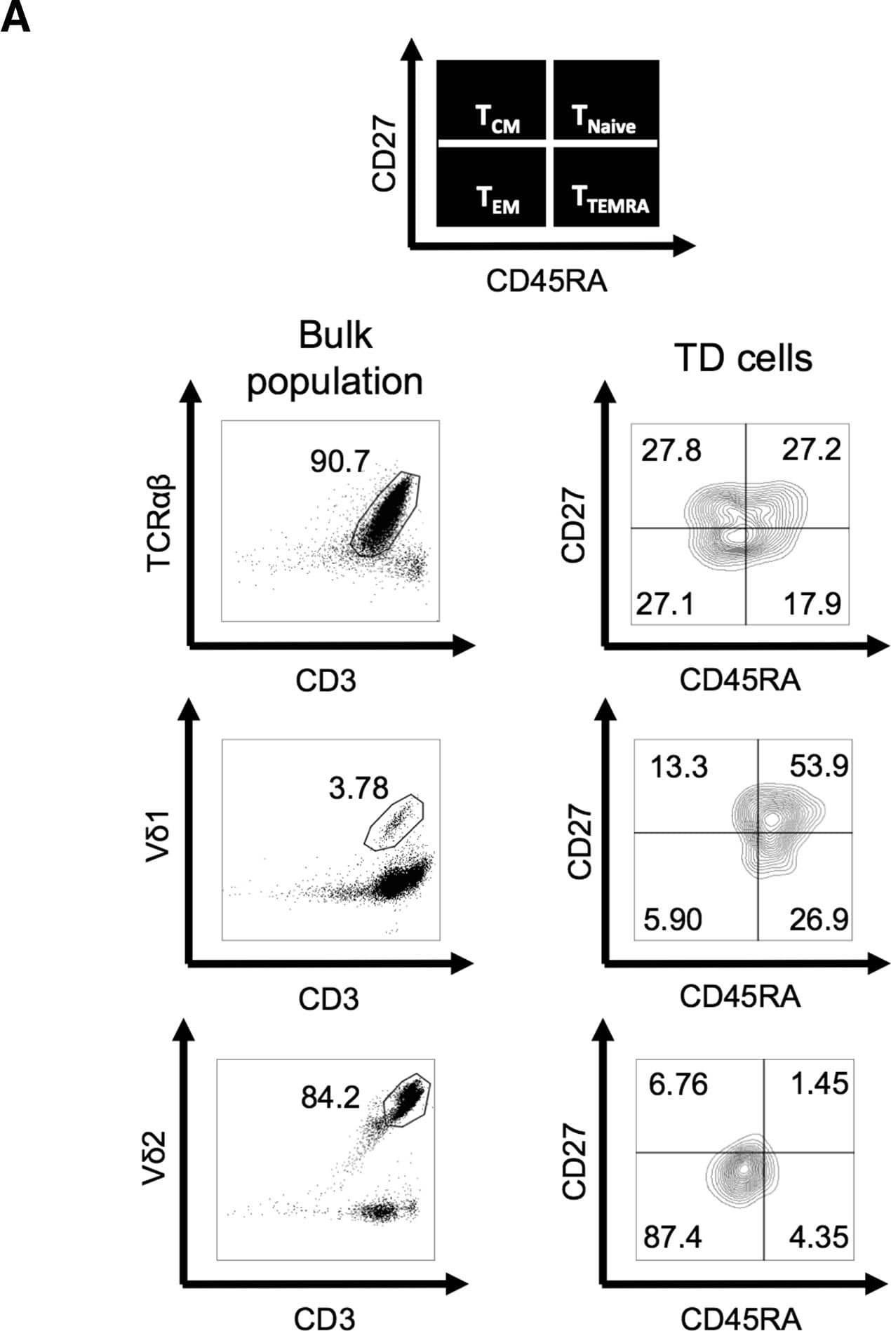
Detection of Human CD34 by Flow Cytometry Memory Phenotype and Exhaustion Marker Expression on Expanded CD3+ alpha beta, Vδ1+, and Vδ2+ Live T Cells(A) Representative flow cytometry dot plot showing gating of T cell subtypes ( alpha beta, Vδ1+, and Vδ2+) and contour plots of the corresponding subtype memory phenotype (N, naive; CM, central memory; EM, effector memory; TEMRA, terminally differentiated effector memory) according to expression of CD27 and CD45RA. (B–D) memory phenotypes of pre-expanded PBMCs (B), post-expansion non-transduced (NTD) cells (C), and post-expansion GD2-CAR-transduced cells (D). alpha beta cells were stimulated with CD3/CD28 antibody, Vδ1 cells were stimulated with ConA, and Vδ2 cells were stimulated with ZOL (data represented as mean ± SEM; alpha beta and Vδ2, n = 6; Vδ1, n = 3). (E and F) Expanded Vδ1+ cells transduced with CAR after ConA stimulation express significantly fewer exhaustion markers (PD1 and TIM3) than alpha beta and Vδ2+ CAR-T cells. (E) Flow cytometry plots from a representative donor. alpha beta cells were stimulated with CD3/CD28 antibody, Vδ1+ cells were stimulated with ConA, and Vδ2+ cells were stimulated with ZOL. (F) PD1 and TIM3 expression on TD and NTD T cells 13 days following activation (data represented as mean ± SEM; alpha beta and Vδ2+, n = 6; Vδ1+, n = 3; p value for significance comparing double-negative populations). Image collected and cropped by CiteAb from the following open publication (https://pubmed.ncbi.nlm.nih.gov/29310916), licensed under a CC-BY license. Not internally tested by R&D Systems.
Reconstitution Calculator
Preparation and Storage
- 12 months from date of receipt, 2 to 8 °C as supplied.
Background: CD34
The CD34 family of cell surface transmembrane proteins includes the hematopoietic progenitor cell antigen CD34, Podocalyxin, and Endoglycan. This single-pass sialomucin‑like transmembrane protein is heavily glycosylated and phosphorylated by Protein kinase C (PKC). CD34 is a 115 kDa glycoprotein found on multipotent precursors, bone marrow stromal cells, embryonic fibroblasts, vascular endothelia, as well as some populations of mesenchymal stem cells, and tumor cell lines. CD34 is involved in the adhesion of stem cells to the bone marrow extracellular matrix or to stromal cells.
Product Datasheets
Citations for Human CD34 APC-conjugated Antibody
R&D Systems personnel manually curate a database that contains references using R&D Systems products. The data collected includes not only links to publications in PubMed, but also provides information about sample types, species, and experimental conditions.
17
Citations: Showing 1 - 10
Filter your results:
Filter by:
-
Tumor infiltrating lymphocytes expanded from pediatric neuroblastoma display heterogeneity of phenotype and function
Authors: Marina Ollé Hurtado, Jolien Wolbert, Jonathan Fisher, Barry Flutter, Sian Stafford, Jack Barton et al.
PLOS ONE
-
Exploration of T cell immune responses by expression of a dominant-negative SHP1 and SHP2
Authors: Julia Taylor, Anna Bulek, Isaac Gannon, Mathew Robson, Evangelia Kokalaki, Thomas Grothier et al.
Frontiers in Immunology
-
Novel Fas-TNFR chimeras that prevent Fas ligand-mediated kill and signal synergistically to enhance CAR T cell efficacy
Authors: Callum McKenzie, Mohamed El-Kholy, Farhaan Parekh, Mathew Robson, Katarina Lamb, Christopher Allen et al.
Molecular Therapy - Nucleic Acids
-
Functional avidity of anti-B7H3 CAR-T constructs predicts antigen density thresholds for triggering effector function
Authors: Barisa, M;Muller, HP;Zappa, E;Shah, R;Buhl, JL;Draper, B;Himsworth, C;Bowers, C;Munnings-Tomes, S;Nicolaidou, M;Morlando, S;Birley, K;Leboreiro-Babe, C;Vitali, A;Privitera, L;O'Sullivan, K;Greppi, A;Buschhaus, M;Román, MB;de Blank, S;van den Ham, F;van 't Veld, BR;Ferry, G;Fisher, J;Shome, D;Nadafi, R;Ansari, IH;Reijmers, R;Giuliani, S;Sondel, P;Donovan, LK;Chesler, L;Jan Molenaar, ;Drost, J;Rios, AC;Chester, K;Wienke, J;Anderson, J;
Nature communications
Species: Human
Sample Types: Whole Cells
Applications: Flow Cytometry, Immunocytochemistry -
TRBC1-CAR T cell therapy in peripheral T cell lymphoma: a phase 1/2 trial
Authors: Cwynarski, K;Iacoboni, G;Tholouli, E;Menne, T;Irvine, DA;Balasubramaniam, N;Wood, L;Shang, J;Xue, E;Zhang, Y;Basilico, S;Neves, M;Raymond, M;Scott, I;El-Kholy, M;Jha, R;Dainton-Smith, H;Hussain, R;Day, W;Ferrari, M;Thomas, S;Lilova, K;Brugger, W;Marafioti, T;Lao-Sirieix, P;Maciocia, P;Pule, M;
Nature medicine
Species: Human
Sample Types: Whole Cells
Applications: Flow Cytometry -
Novel Fas-TNFR chimeras that prevent Fas ligand-mediated kill and signal synergistically to enhance CAR T cell efficacy
Authors: Callum McKenzie, Mohamed El-Kholy, Farhaan Parekh, Mathew Robson, Katarina Lamb, Christopher Allen et al.
Molecular Therapy - Nucleic Acids
Species: Human
Sample Types: Whole Cells
Applications: Flow Cytometry -
Role of endothelial colony forming cells (ECFCs) Tetrahydrobiopterin (BH4) in determining ECFCs functionality in coronary artery disease (CAD) patients
Authors: A Sen, A Singh, A Roy, S Mohanty, N Naik, M Kalaivani, L Ramakrishn
Scientific Reports, 2022-02-23;12(1):3076.
Species: Human
Sample Types: Whole Cells
Applications: Flow Cytometry -
Different concentrations of C5a affect human dental pulp mesenchymal stem cells differentiation
Authors: J Liu, X Wei, J Hu, X Tan, X Kang, L Gao, N Li, X Shi, M Yuan, W Hu, M Liu
BMC Oral Health, 2021-09-24;21(1):470.
Species: Human
Sample Types: Whole Cells
Applications: Flow Cytometry -
Targeted multi-epitope switching enables straightforward positive/negative selection of CAR T cells
Authors: L Mosti, LM Langner, KO Chmielewsk, P Arbuthnot, J Alzubi, T Cathomen
Gene Therapy, 2021-02-01;0(0):.
Species: Human
Sample Types: Whole Cells
Applications: Flow Cytometry -
Identification and Targeting of Thomsen-Friedenreich and IL1RAP Antigens on Chronic Myeloid Leukemia Stem Cells Using Bi-Specific Antibodies
Authors: RE Eldesouki, C Wu, FM Saleh, EA Mohammed, S Younes, NE Hassan, TC Brown, EU Alt, JE Robinson, FM Badr, SE Braun
OncoTargets and Therapy, 2021-01-22;14(0):609-621.
Species: Human
Sample Types: Whole Cells
Applications: Flow Cytometry -
Repurposing endogenous immune pathways to tailor and control chimeric antigen receptor T�cell functionality
Authors: M Sachdeva, BW Busser, S Temburni, B Jahangiri, AS Gautron, A Maréchal, A Juillerat, A Williams, S Depil, P Duchateau, L Poirot, J Valton
Nat Commun, 2019-11-13;10(1):5100.
Species: Human
Sample Types: Whole Cells
Applications: Flow Cytometry -
Tumor infiltrating lymphocytes expanded from pediatric neuroblastoma display heterogeneity of phenotype and function
Authors: Marina Ollé Hurtado, Jolien Wolbert, Jonathan Fisher, Barry Flutter, Sian Stafford, Jack Barton et al.
PLOS ONE
Species: Human
Sample Types: Whole Cells
Applications: Flow Cytometry -
Constitutively active MyD88/CD40 costimulation enhances expansion and efficacy of chimeric antigen receptor T cells targeting hematological malignancies
Authors: MR Collinson-, WC Chang, A Lu, M Khalil, JW Crisostomo, PY Lin, A Mahendrava, NP Shinners, ME Brandt, M Zhang, M Duong, JH Bayle, KM Slawin, DM Spencer, AE Foster
Leukemia, 2019-02-28;0(0):.
Species: Human
Sample Types: Whole Cells
Applications: CAR-T (Flow Cytometry) -
Enhanced Anti-lymphoma Activity of CAR19-iNKT Cells Underpinned by Dual CD19 and CD1d Targeting
Authors: Antonia Rotolo, Valentina S. Caputo, Monika Holubova, Nicoleta Baxan, Olivier Dubois, Mohammed Suhail Chaudhry et al.
Cancer Cell
Species: Human
Sample Types: Whole Cells
Applications: Flow Cytometry -
A Versatile Safeguard for Chimeric Antigen Receptor T-Cell Immunotherapies
Authors: J Valton, V Guyot, B Boldajipou, C Sommer, T Pertel, A Juillerat, A Duclert, BJ Sasu, P Duchateau, L Poirot
Sci Rep, 2018-06-12;8(1):8972.
Species: Human
Sample Types: Whole Cells
Applications: Flow Cytometry -
Chimeric Antigen Receptor-Engineered Human Gamma Delta T Cells: Enhanced Cytotoxicity with Retention of Cross Presentation
Authors: A Capsomidis, G Benthall, HH Van Acker, J Fisher, AM Kramer, Z Abeln, Y Majani, T Gileadi, R Wallace, K Gustafsson, B Flutter, J Anderson
Mol. Ther., 2017-12-08;0(0):.
Species: Human
Sample Types: Whole Cells
Applications: Flow Cytometry -
Human theca arises from ovarian stroma and is comprised of three discrete subtypes
Authors: Nicole Lustgarten Guahmich, Limor Man, Jerry Wang, Laury Arazi, Eleni Kallinos, Ariana Topper-Kroog et al.
Communications Biology
FAQs
No product specific FAQs exist for this product, however you may
View all Antibody FAQsReviews for Human CD34 APC-conjugated Antibody
There are currently no reviews for this product. Be the first to review Human CD34 APC-conjugated Antibody and earn rewards!
Have you used Human CD34 APC-conjugated Antibody?
Submit a review and receive an Amazon gift card.
$25/€18/£15/$25CAN/¥75 Yuan/¥2500 Yen for a review with an image
$10/€7/£6/$10 CAD/¥70 Yuan/¥1110 Yen for a review without an image


