ErbB2, also called Neu and Her2 (human epidermal growth factor receptor 2), is a type I membrane glycoprotein that is a member of the ErbB family of tyrosine kinase receptors. ErbB family members serve as receptors for the epidermal growth factor (EGF) family of growth factors. ErbB2 is widely expressed in epithelial cells and has also been found to be over-expressed in a large number of breast carcinomas. Among ErbB family members, ErbB2 is unique in that it has no identified ligands. Rather, ErbB2 heterodimerizes with the other members of the ErbB family (ErbB1 (EGF R), ErbB3, ErbB4) to form higher affinity signaling complexes. Because ErbB3 contains a defective kinase domain, the kinase domain of ErbB2 is responsible for initiating the tyrosine phosphorylation signal through the heterodimeric receptor. It has been found that a discrete three amino acid signal in the ErbB3 cytoplasmic domain is critical for transactivation of ErbB2. Interestingly, this same three amino acid signal has also been found in ErbB1 and ErbB4. Phosphoinositide 3-kinase has been shown to play a role in ErbB2 signal transduction. The cytoplasmic domain of ErbB2 has been shown to associate with beta-catenin and plakoglobin. Human ErbB2 consists of 1255 amino acids (aa) with a 21 aa signal sequence, a 631 aa extracellular domain, a 23 aa transmembrane region, and a 580 aa cytoplasmic domain. ErbB2 can be shed from the cell surface by proteolytic cleavage by an unidentified protease. ErbB2 appears to play roles in development, cancer, communication at the neuromuscular junction and regulation of cell growth and differentiation (1-10).
Human ErbB2/Her2 Biotinylated Antibody
R&D Systems | Catalog # BAF1129

Key Product Details
Species Reactivity
Validated:
Cited:
Applications
Validated:
Cited:
Label
Antibody Source
Product Specifications
Immunogen
Thr23-Thr652
Accession # P04626
Specificity
Clonality
Host
Isotype
Scientific Data Images for Human ErbB2/Her2 Biotinylated Antibody
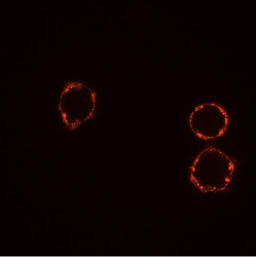
ErbB2/Her2 in MCF-7 Human Cell Line.
ErbB2/Her2 was detected in immersion fixed MCF-7 human breast cancer cell line using Goat Anti-Human ErbB2/Her2 Biotinylated Antigen Affinity-purified Polyclonal Antibody (Catalog # BAF1129) at 10 µg/mL for 3 hours at room temperature. Cells were stained using the NorthernLights™ 557-conjugated Streptavidin (yellow; Catalog # NL999) and counterstained with DAPI (blue). View our protocol for Fluorescent ICC Staining of Cells on Coverslips.

ErbB2/Her2 in Human Breast Cancer Tissue.
ErbB2/Her2 was detected in immersion fixed paraffin-embedded sections of human breast cancer tissue using Goat Anti-Human ErbB2/Her2 Biotinylated Antigen Affinity-purified Polyclonal Antibody (Catalog # BAF1129) at 15 µg/mL overnight at 4 °C. Tissue was stained using the Anti-Goat HRP-DAB Cell & Tissue Staining Kit (brown; Catalog # CTS008) and counterstained with hematoxylin (blue). Specific staining was localized to plasma membrane. View our protocol for Chromogenic IHC Staining of Paraffin-embedded Tissue Sections.
Applications for Human ErbB2/Her2 Biotinylated Antibody
Immunocytochemistry
Sample: Immersion fixed MCF-7 human breast cancer cell line
Immunohistochemistry
Sample: Immersion fixed paraffin-embedded sections of human breast cancer tissue
Western Blot
Sample: Recombinant Human ErbB2/Her2 Fc Chimera (Catalog # 1129-ER)
Human ErbB2/Her2 Sandwich Immunoassay
Reviewed Applications
Read 1 review rated 5 using BAF1129 in the following applications:
Formulation, Preparation, and Storage
Purification
Reconstitution
Reconstitute at 0.2 mg/mL in sterile PBS.
Formulation
Shipping
Stability & Storage
- 12 months from date of receipt, -20 to -70 °C as supplied.
- 1 month, 2 to 8 °C under sterile conditions after reconstitution.
- 6 months, -20 to -70 °C under sterile conditions after reconstitution.
Calculators
Background: ErbB2/Her2
References
- Coussens, L. et al. (1985) Science 230:1132.
- Yamamoto, T. et al. (1986) Nature 319:230.
- Kanai, Y. et al. (1995) Biochem. Biophys. Res. Commun. 208:1067.
- Codony-Servat, J. et al. (1999) Cancer Res. 59:1196.
- Carraway, K.L. 3rd et al. (1994) J. Biol. Chem. 269:14303.
- Emkey, R. and C.R. Kahn (1997) J. Biol. Chem. 272:31172.
- Schaefer, G. et al. (1999) J. Biol. Chem. 274:859.
- Schlessinger, J. (2000) Cell 103:211.
- Hellyer, N.J. et al. (2001) J. Biol. Chem. 276:42153.
- Daly, R.J. (1999) Growth Factors 16:255.
Long Name
Alternate Names
Gene Symbol
UniProt
Additional ErbB2/Her2 Products
Product Documents for Human ErbB2/Her2 Biotinylated Antibody
Certificate of Analysis
To download a Certificate of Analysis, please enter a lot or batch number in the search box below.
Note: Certificate of Analysis not available for kit components.
Product Specific Notices for Human ErbB2/Her2 Biotinylated Antibody
For research use only
Related Research Areas
Citations for Human ErbB2/Her2 Biotinylated Antibody
 Powered by Bioz
Customer Reviews for Human ErbB2/Her2 Biotinylated Antibody (1)
Have you used Human ErbB2/Her2 Biotinylated Antibody?
Submit a review and receive an Amazon gift card!
$25/€18/£15/$25CAN/¥2500 Yen for a review with an image
$10/€7/£6/$10CAN/¥1110 Yen for a review without an image
Submit a review
Customer Images
-
Application: Immunocytochemistry/ImmunofluorescenceSample Tested: SK-BR-3 human breast cancer cell lineSpecies: GoatVerified Customer | Posted 02/03/2016SK-BR-3 cell staining with Biotin-labeled pAb (BAF1129) and Streptavidin, Alexa Fluor® 633 conjugate.

There are no reviews that match your criteria.
Protocols
Find general support by application which include: protocols, troubleshooting, illustrated assays, videos and webinars.
- Antigen Retrieval Protocol (PIER)
- Antigen Retrieval for Frozen Sections Protocol
- Appropriate Fixation of IHC/ICC Samples
- Cellular Response to Hypoxia Protocols
- Chromogenic IHC Staining of Formalin-Fixed Paraffin-Embedded (FFPE) Tissue Protocol
- Chromogenic Immunohistochemistry Staining of Frozen Tissue
- ClariTSA™ Fluorophore Kits
- Detection & Visualization of Antibody Binding
- Fluorescent IHC Staining of Frozen Tissue Protocol
- Graphic Protocol for Heat-induced Epitope Retrieval
- Graphic Protocol for the Preparation and Fluorescent IHC Staining of Frozen Tissue Sections
- Graphic Protocol for the Preparation and Fluorescent IHC Staining of Paraffin-embedded Tissue Sections
- Graphic Protocol for the Preparation of Gelatin-coated Slides for Histological Tissue Sections
- ICC Cell Smear Protocol for Suspension Cells
- ICC Immunocytochemistry Protocol Videos
- ICC for Adherent Cells
- IHC Sample Preparation (Frozen sections vs Paraffin)
- Immunocytochemistry (ICC) Protocol
- Immunocytochemistry Troubleshooting
- Immunofluorescence of Organoids Embedded in Cultrex Basement Membrane Extract
- Immunofluorescent IHC Staining of Formalin-Fixed Paraffin-Embedded (FFPE) Tissue Protocol
- Immunohistochemistry (IHC) and Immunocytochemistry (ICC) Protocols
- Immunohistochemistry Frozen Troubleshooting
- Immunohistochemistry Paraffin Troubleshooting
- Preparing Samples for IHC/ICC Experiments
- Preventing Non-Specific Staining (Non-Specific Binding)
- Primary Antibody Selection & Optimization
- Protocol for Heat-Induced Epitope Retrieval (HIER)
- Protocol for Making a 4% Formaldehyde Solution in PBS
- Protocol for VisUCyte™ HRP Polymer Detection Reagent
- Protocol for the Fluorescent ICC Staining of Cell Smears - Graphic
- Protocol for the Fluorescent ICC Staining of Cultured Cells on Coverslips - Graphic
- Protocol for the Preparation & Fixation of Cells on Coverslips
- Protocol for the Preparation and Chromogenic IHC Staining of Frozen Tissue Sections
- Protocol for the Preparation and Chromogenic IHC Staining of Frozen Tissue Sections - Graphic
- Protocol for the Preparation and Chromogenic IHC Staining of Paraffin-embedded Tissue Sections
- Protocol for the Preparation and Chromogenic IHC Staining of Paraffin-embedded Tissue Sections - Graphic
- Protocol for the Preparation and Fluorescent ICC Staining of Cells on Coverslips
- Protocol for the Preparation and Fluorescent ICC Staining of Non-adherent Cells
- Protocol for the Preparation and Fluorescent ICC Staining of Stem Cells on Coverslips
- Protocol for the Preparation and Fluorescent IHC Staining of Frozen Tissue Sections
- Protocol for the Preparation and Fluorescent IHC Staining of Paraffin-embedded Tissue Sections
- Protocol for the Preparation of Gelatin-coated Slides for Histological Tissue Sections
- Protocol for the Preparation of a Cell Smear for Non-adherent Cell ICC - Graphic
- R&D Systems Quality Control Western Blot Protocol
- TUNEL and Active Caspase-3 Detection by IHC/ICC Protocol
- The Importance of IHC/ICC Controls
- Troubleshooting Guide: Immunohistochemistry
- Troubleshooting Guide: Western Blot Figures
- Western Blot Conditions
- Western Blot Protocol
- Western Blot Protocol for Cell Lysates
- Western Blot Troubleshooting
- Western Blot Troubleshooting Guide
- View all Protocols, Troubleshooting, Illustrated assays and Webinars


