Human ER beta /NR3A2 Antibody Summary
Met1-Gly318
Accession # Q92731
Applications
Please Note: Optimal dilutions should be determined by each laboratory for each application. General Protocols are available in the Technical Information section on our website.
Scientific Data
 View Larger
Detection of Human ER beta /NR3A2 by Western Blot. Western blot shows lysates of MCF-7 human breast cancer cell line, Saos-2 human osteosarcoma cell line, and T47D human breast cancer cell line. PVDF membrane was probed with 2 µg/mL of Mouse Anti-Human ER beta /NR3A2 Monoclonal Antibody (Catalog # MAB7106) followed by HRP-conjugated Anti-Mouse IgG Secondary Antibody (Catalog # HAF007). A specific band was detected for ER beta /NR3A2 at approximately 48 kDa (as indicated). This experiment was conducted under reducing conditions and using Immunoblot Buffer Group 1.
 View Larger
ER beta /NR3A2 in LNCaP Human Cell Line. ER beta /NR3A2 was detected in immersion fixed LNCaP human prostate cancer cell line using Mouse Anti-Human ER beta /NR3A2 Monoclonal Antibody (Catalog # MAB7106) at 10 µg/mL for 3 hours at room temperature. Cells were stained using the NorthernLights™ 557-conjugated Anti-Mouse IgG Secondary Antibody (red; Catalog # NL007) and counter-stained with DAPI (blue). Specific staining was localized to cytoplasm. View our protocol for Fluorescent ICC Staining of Cells on Coverslips.
 View Larger
ER beta /NR3A2 in MCF‑7 Human Cell Line. ER beta /NR3A2 was detected in immersion fixed MCF-7 human breast cancer cell line using Mouse Anti-Human ER beta /NR3A2 Monoclonal Antibody (Catalog # MAB7106) at 10 µg/mL for 3 hours at room temperature. Cells were stained using the NorthernLights™ 557-conjugated Anti-Mouse IgG Secondary Antibody (red; Catalog # NL007) and counter-stained with DAPI (blue). Specific staining was localized to cytoplasm and nuclei. View our protocol for Fluorescent ICC Staining of Cells on Coverslips.
 View Larger
Detection of ER beta /NR3A2 in MCF‑7 Human Cell Line by Flow Cytometry. MCF-7 human breast cancer cell line was stained with Mouse Anti-Human ER beta /NR3A2 Monoclonal Antibody (Catalog # MAB7106, filled histogram) or isotype control antibody (Catalog # MAB002, open histogram), followed by Phycoerythrin-conjugated Anti-Mouse IgG Secondary Antibody (Catalog # F0102B). To facilitate intracellular staining, cells were fixed with paraformaldehyde and permeabilized with saponin.
 View Larger
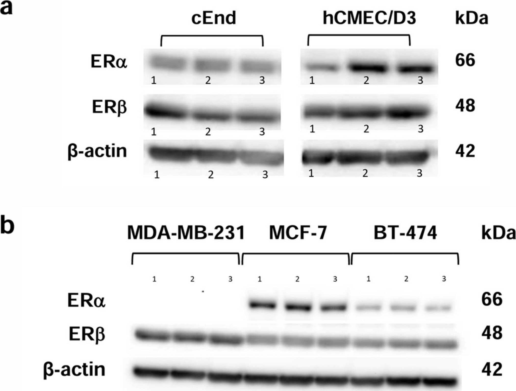
Detection of ER beta /NR3A2 by Western Blot Western blot analysis showing the protein expression patterns of the estrogen receptors (ERs) ER alpha (66 kDa) and ER beta (48 kDa). (a) The murine brain endothelial cell lines cEND (left) and the human brain endothelial cell line hCMEC/D3 (right) show the presence of both ERs in three experimental runs (1, 2, 3). (b) The breast cancer (BC) cell lines showed the presence of ER beta in MCF-7, BT-474 and MDA-MB-231 cells, whereas ER alpha was only detected in MCF-7 and BT-474. Again, three experimental runs were performed (1, 2, 3). beta -actin (42 kDa) was used as a loading control. Image collected and cropped by CiteAb from the following open publication (https://www.mdpi.com/1422-0067/25/6/3379), licensed under a CC-BY license. Not internally tested by R&D Systems.
 View Larger
Detection of Human and Mouse ER beta /NR3A2 by Simple WesternTM. Simple Western lane view shows lysates of MCF‑7 human breast cancer cell line, T47D human breast cancer cell line, MDA‑MB‑231 human breast cancer cell line, and 3T3‑L1 mouse embryonic fibroblast adipose-like cell line, loaded at 0.2 mg/mL. A specific band was detected for ER beta /NR3A2 at approximately 54 kDa (as indicated) using 50 µg/mL of Mouse Anti-Human ER beta /NR3A2 Monoclonal Antibody (Catalog # MAB7106). This experiment was conducted under reducing conditions and using the 12-230 kDa separation system.
Reconstitution Calculator
Preparation and Storage
- 12 months from date of receipt, -20 to -70 °C as supplied.
- 1 month, 2 to 8 °C under sterile conditions after reconstitution.
- 6 months, -20 to -70 °C under sterile conditions after reconstitution.
Background: ER beta/NR3A2
Estrogen Receptor beta (ER beta ; NR3A2) is a member of the steroid receptor family. The natural ligand for ER is the classical estrogenic compound 17 beta -estradiol. ER beta is expressed in the granulosa cell layer of primary, secondary and mature follicles in the ovary, in bone, bladder, uterus, testis, epididymis, gastrointestinal tract, kidney, breast, heart, vessel wall, immune system, lung, pituitary, hippocampus and hypothalamus. Roles for ER beta in the reproductive and cardiovascular systems have been reported, although these are the subject of conflicting reports. ER beta has been postulated to act primarily as a modulator of ER alpha function. ER beta has been shown to form homodimers as well as heterodimers with ER alpha. Both ER alpha and ER beta can give rise to numerous isoforms.
Product Datasheets
Citations for Human ER beta /NR3A2 Antibody
R&D Systems personnel manually curate a database that contains references using R&D Systems products. The data collected includes not only links to publications in PubMed, but also provides information about sample types, species, and experimental conditions.
3
Citations: Showing 1 - 3
Filter your results:
Filter by:
-
Genes Co-Expressed with ESR2 Influence Clinical Outcomes in Cancer Patients: TCGA Data Analysis
Authors: Lipowicz, JM;Mali?ska, A;Nowicki, M;Raw?uszko-Wieczorek, AA;
International journal of molecular sciences
Species: Human
Sample Types: Whole Tissue
Applications: Immunohistochemistry -
Time since menopause and skeletal muscle estrogen receptors, PGC-1 alpha, and AMPK
Authors: Young-Min Park, Rocio I. Pereira, Christopher B. Erickson, Tracy A. Swibas, Chounghun Kang, Rachael E. Van Pelt
Menopause
-
Estradiol-mediated improvements in adipose tissue insulin sensitivity are related to the balance of adipose tissue estrogen receptor ? and ? in postmenopausal women
Authors: YM Park, RI Pereira, CB Erickson, TA Swibas, KA Cox-York, RE Van Pelt
PLoS ONE, 2017-05-04;12(5):e0176446.
Species: Human
Sample Types: Cell Lysates
Applications: Western Blot
FAQs
No product specific FAQs exist for this product, however you may
View all Antibody FAQsReviews for Human ER beta /NR3A2 Antibody
Average Rating: 4.5 (Based on 2 Reviews)
Have you used Human ER beta /NR3A2 Antibody?
Submit a review and receive an Amazon gift card.
$25/€18/£15/$25CAN/¥75 Yuan/¥2500 Yen for a review with an image
$10/€7/£6/$10 CAD/¥70 Yuan/¥1110 Yen for a review without an image
Filter by:


