Human FABP5/E-FABP Antibody Summary
Ala2-Glu135
Accession # Q01469
*Small pack size (-SP) is supplied either lyophilized or as a 0.2 µm filtered solution in PBS.
Applications
Please Note: Optimal dilutions should be determined by each laboratory for each application. General Protocols are available in the Technical Information section on our website.
Scientific Data
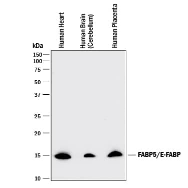 View Larger
Detection of Human FABP5/E‑FABP by Western Blot. Western blot shows lysates of human heart tissue, human brain (cerebellum) tissue, and human placenta tissue. PVDF membrane was probed with 1 µg/mL of Goat Anti-Human FABP5/E-FABP Antigen Affinity-purified Polyclonal Antibody (Catalog # AF3077) followed by HRP-conjugated Anti-Goat IgG Secondary Antibody (Catalog # HAF017). A specific band was detected for FABP5/E-FABP at approximately 15 kDa (as indicated). This experiment was conducted under reducing conditions and using Immunoblot Buffer Group 1.
 View Larger
Detection of Human FABP5/E‑FABP by Simple WesternTM. Simple Western lane view shows lysates of Exosome Standards (Human Urine) (NBP2-49840) and human placenta tissue, loaded at 0.5 mg/ml. A specific band was detected for FABP5/E‑FABP at approximately 20 kDa (as indicated) using 10 µg/ml of Goat Anti-Human FABP5/E‑FABP Antigen Affinity-purified Polyclonal Antibody (Catalog # AF3077) followed by HRP-conjugated Donkey Anti-Goat Secondary Antibody (Catalog # 042-206). This experiment was conducted under reducing conditions and using the 2-40kDa separation system.
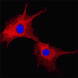 View Larger
FABP5/E‑FABP in HUVEC Human Umbilical Vein Endothelial Cells. FABP5/E-FABP was detected in immersion fixed HUVEC human umbilical vein endothelial cells using Goat Anti-Human FABP5/E-FABP Antigen Affinity-purified Polyclonal Antibody (Catalog # AF3077) at 10 µg/mL for 3 hours at room temperature. Cells were stained using the NorthernLights™ 557-conjugated Anti-Goat IgG Secondary Antibody (red; Catalog # NL001) and counterstained with DAPI (blue). Specific staining was localized to cytoplasm. View our protocol for Fluorescent ICC Staining of Non-adherent Cells.
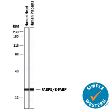 View Larger
Detection of Human FABP5/E‑FABP by Simple WesternTM. Simple Western lane view shows lysates of human heart tissue and human placenta tissue, loaded at 0.2 mg/mL. A specific band was detected for FABP5/E-FABP at approximately 21 kDa (as indicated) using 10 µg/mL of Goat Anti-Human FABP5/E-FABP Antigen Affinity-purified Polyclonal Antibody (Catalog # AF3077) followed by 1:50 dilution of HRP-conjugated Anti-Goat IgG Secondary Antibody (Catalog # HAF109). This experiment was conducted under reducing conditions and using the 12-230 kDa separation system.
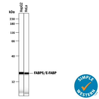 View Larger
Detection of Human FABP5/E‑FABP by Simple WesternTM. Simple Western lane view shows lysates of HepG2 human hepatocellular carcinoma cell line and HeLa human cervical epithelial carcinoma cell line, loaded at 0.2 mg/mL. A specific band was detected for FABP5/E-FABP at approximately 21 kDa (as indicated) using 12.5 µg/mL of Goat Anti-Human FABP5/E-FABP Antigen Affinity-purified Polyclonal Antibody (Catalog # AF3077) followed by 1:50 dilution of HRP-conjugated Anti-Goat IgG Secondary Antibody (Catalog # HAF109). This experiment was conducted under reducing conditions and using the 12-230 kDa separation system.
 View Larger
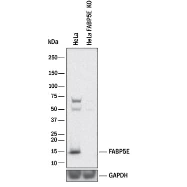
Western Blot Shows Human FABP5/E‑FABP Specificity by Using Knockout Cell Line. Western blot shows lysates of HeLa human cervical epithelial carcinoma parental cell line and FABP5/E-FABP knockout HeLa cell line (KO). PVDF membrane was probed with 1 µg/mL of Goat Anti-Human FABP5/E-FABP Antigen Affinity-purified Polyclonal Antibody (Catalog # AF3077) followed by HRP-conjugated Anti-Goat IgG Secondary Antibody (Catalog # HAF017). A specific band was detected for FABP5/E-FABP at approximately 15 kDa (as indicated) in the parental HeLa cell line, but is not detectable in knockout HeLa cell line. GAPDH (Catalog # AF5718) is shown as a loading control. This experiment was conducted under reducing conditions and using Immunoblot Buffer Group 1.
 View Larger
Detection of Mouse FABP5/E-FABP by Western Blot Rotenone induced neurotoxicity, especially with co-overexpression of FABP5 and alpha -Synuclein. (A) Representative images of immunoblots probed with antibodies against alpha Syn (~19 kDa) and FABP5 (~12 kDa). Blots with anti-beta -tubulin antibody showed that a similar amount of protein was loaded. (B,C) Viability of Neuro-2A cells treated with indicated concentrations of rotenone for 48 h was assessed using CCK-8 assays. The transfection conditions were pcDNA 3.1-transfected cells (mock) and FABP5-transfected cells in (B); alpha Syn-transfected cells and alpha Syn/FABP5-transfected cells ( alpha S/F5) in (C). Results are presented as means ± SEM (n = 4 parallel cell experiments). * p < 0.05, ** p < 0.01, vs. 0 μM rotenone groups; ## p < 0.01 vs. alpha Syn-transfected cells. CCK-8: cell counting kit-8. Image collected and cropped by CiteAb from the following open publication (https://pubmed.ncbi.nlm.nih.gov/33499263), licensed under a CC-BY license. Not internally tested by R&D Systems.
 View Larger
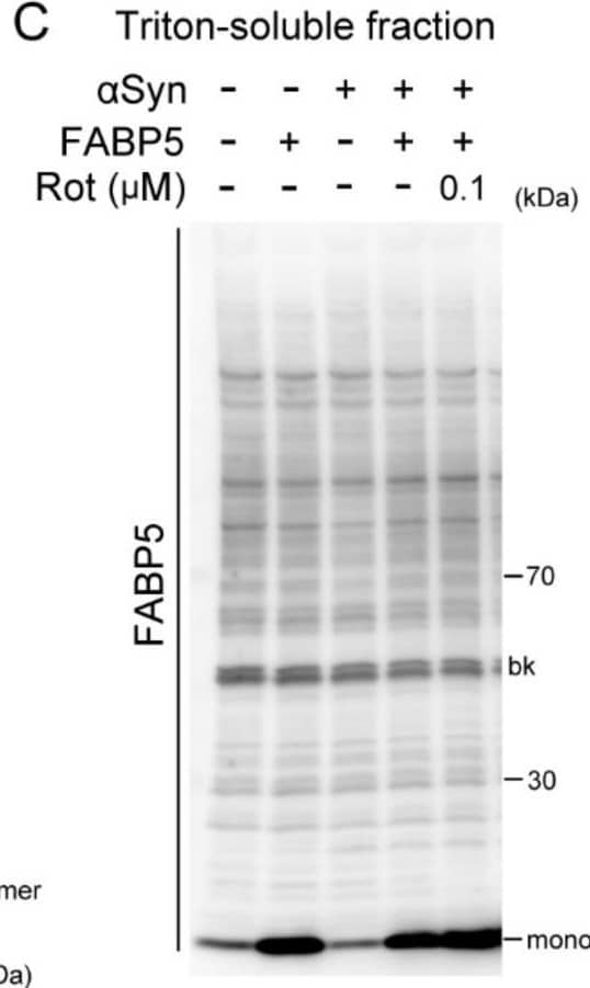
Detection of Mouse FABP5/E-FABP by Western Blot Effects of rotenone on alpha Syn oligomerization and aggregation in FABP5/ alpha Syn-transfected Neuro-2A cells. (A–D) Representative images of immunoblots probed with antibodies against alpha Syn and FABP5 under non-denaturing conditions of Triton-soluble and SDS-soluble fractions. Detection with anti-beta -tubulin antibody was used as a protein-loading control. (E–G) Quantitative analyses of protein contents of different molecular weights, which indicate protein complexes of dimer/trimers, oligomers, and aggregates. Results are presented as means ± SEM (n = 3 respective cell experiments). * p < 0.05, ** p < 0.01, vs. 0.1 μM rotenone-treated alpha S/F5 cells. n.s., not statistically significant. D/T: dimers/trimers; O: oligomers; A: aggregates. bk besides blots indicate non-specific bands. Image collected and cropped by CiteAb from the following open publication (https://pubmed.ncbi.nlm.nih.gov/33499263), licensed under a CC-BY license. Not internally tested by R&D Systems.
Reconstitution Calculator
Preparation and Storage
- 12 months from date of receipt, -20 to -70 °C as supplied.
- 1 month, 2 to 8 °C under sterile conditions after reconstitution.
- 6 months, -20 to -70 °C under sterile conditions after reconstitution.
Background: FABP5/E-FABP
Human FABP-5, also known as epidermal fatty acid binding protein (E-FABP), is a 15 kDa member of a cytosolic fatty acid binding protein superfamily. It is associated with keratinocytes and adipocytes and is suggested to promote fatty acid availability to enzymes, protect cell structures from fatty acid attack, and target fatty acids to nuclear transcription factors. The amino acid sequence of human FABP5 is 80%, 81% and 92% identical to that of mouse, rat and bovine FABP5, respectively.
Product Datasheets
Citations for Human FABP5/E-FABP Antibody
R&D Systems personnel manually curate a database that contains references using R&D Systems products. The data collected includes not only links to publications in PubMed, but also provides information about sample types, species, and experimental conditions.
12
Citations: Showing 1 - 10
Filter your results:
Filter by:
-
Genetic Ablation of the Fatty Acid–Binding Protein FABP5 Suppresses HER2-Induced Mammary Tumorigenesis
Authors: Liraz Levi, Glenn Lobo, Mary Kathryn Doud, Johannes von Lintig, Darcie Seachrist, Gregory P. Tochtrop et al.
Cancer Research
-
Symbiotic Macrophage-Glioma Cell Interactions Reveal Synthetic Lethality in PTEN-Null Glioma
Authors: Chen P, Zhao D, Li J et al.
Cancer Cell
-
A novel fatty acid-binding protein 5 and 7 inhibitor ameliorates oligodendrocyte injury in multiple sclerosis mouse models
Authors: A Cheng, W Jia, I Kawahata, K Fukunaga
EBioMedicine, 2021-10-05;72(0):103582.
Species: Mouse
Sample Types: Tissue Lysates
Applications: Western Blot -
Fatty Acid-Binding Proteins Aggravate Cerebral Ischemia-Reperfusion Injury in Mice
Authors: Q Guo, I Kawahata, T Degawa, Y Ikeda-Mats, M Sun, F Han, K Fukunaga
Biomedicines, 2021-05-10;9(5):.
Species: Mouse
Sample Types: Tissue Homogenates
Applications: Western Blot -
Epidermal Fatty Acid-Binding Protein 5 (FABP5) Involvement in Alpha-Synuclein-Induced Mitochondrial Injury under Oxidative Stress
Authors: Y Wang, Y Shinoda, A Cheng, I Kawahata, K Fukunaga
Biomedicines, 2021-01-22;9(2):.
Species: Human
Sample Types: Cell Lysates, Whole Cells
Applications: ICC, Western Blot -
Fatty Acid Binding Protein 5 Mediates Cell Death by Psychosine Exposure through Mitochondrial Macropores Formation in Oligodendrocytes
Authors: A Cheng, I Kawahata, K Fukunaga
Biomedicines, 2020-12-20;8(12):.
Species: Human
Sample Types: Cell Lysates, Whole Cells
Applications: ICC, Immunoprecipitation, Western Blot -
Curcumin restores sensitivity to retinoic acid in triple negative breast cancer cells.
Authors: Thulasiraman P, McAndrews D, Mohiudddin I
BMC Cancer, 2014-09-27;14(0):724.
Species: Human
Sample Types: Cell Lysates
Applications: Western Blot -
Genetic Ablation of the Fatty Acid–Binding Protein FABP5 Suppresses HER2-Induced Mammary Tumorigenesis
Authors: Liraz Levi, Glenn Lobo, Mary Kathryn Doud, Johannes von Lintig, Darcie Seachrist, Gregory P. Tochtrop et al.
Cancer Research
Species: Human, Mouse
Sample Types: Cell Extracts, Tissue Homogenates
Applications: Western Blot -
Expression of fatty acid-binding proteins in adult hippocampal neurogenic niche of postischemic monkeys.
Authors: Boneva NB, Kaplamadzhiev DB, Sahara S, Kikuchi H, Pyko IV, Kikuchi M, Tonchev AB, Yamashima T
Hippocampus, 2011-02-01;21(2):162-71.
Species: Primate - Macaca fuscata (Japanese Macaque)
Sample Types: Tissue Homogenates, Whole Tissue
Applications: IHC-Fr, Western Blot -
Fatty acid-binding protein 5 and PPARbeta/delta are critical mediators of epidermal growth factor receptor-induced carcinoma cell growth.
Authors: Kannan-Thulasiraman P, Seachrist DD, Mahabeleshwar GH, Jain MK, Noy N
J. Biol. Chem., 2010-04-27;285(25):19106-15.
Species: Human
Sample Types: Cell Lysates
Applications: Western Blot -
Proteogenomic analysis of psoriasis reveals discordant and concordant changes in mRNA and protein abundance
Authors: William R. Swindell, Henriette A. Remmer, Mrinal K. Sarkar, Xianying Xing, Drew H. Barnes, Liza Wolterink et al.
Genome Medicine
-
Saturated fatty acids regulate retinoic acid signalling and suppress tumorigenesis by targeting fatty acid-binding protein 5
Authors: Liraz Levi, Zeneng Wang, Mary Kathryn Doud, Stanley L. Hazen, Noa Noy
Nature Communications
FAQs
No product specific FAQs exist for this product, however you may
View all Antibody FAQsReviews for Human FABP5/E-FABP Antibody
There are currently no reviews for this product. Be the first to review Human FABP5/E-FABP Antibody and earn rewards!
Have you used Human FABP5/E-FABP Antibody?
Submit a review and receive an Amazon gift card.
$25/€18/£15/$25CAN/¥75 Yuan/¥2500 Yen for a review with an image
$10/€7/£6/$10 CAD/¥70 Yuan/¥1110 Yen for a review without an image
