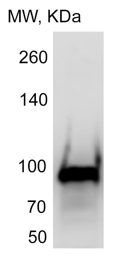
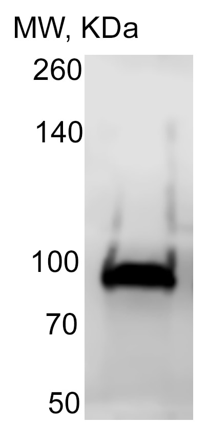
Human gp96/HSP90B1 Antibody Summary
Arg503-Arg660
Accession # P14625
Applications
Please Note: Optimal dilutions should be determined by each laboratory for each application. General Protocols are available in the Technical Information section on our website.
Scientific Data
 View Larger
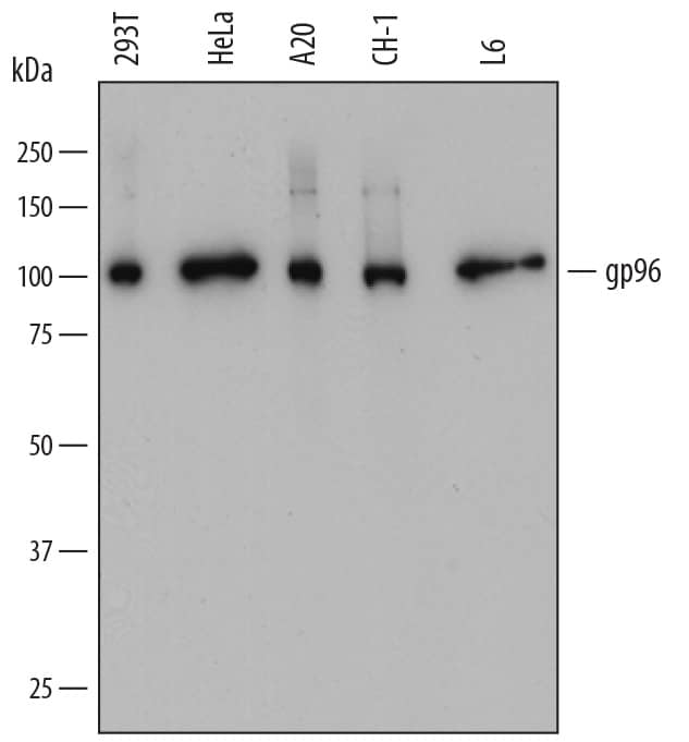
Detection of Human, Mouse, and Rat gp96/HSP90B1 by Western Blot. Western blot shows lysates of 293T human embryonic kidney cell line, HeLa human cervical epithelial carcinoma cell line, A20 mouse B cell lymphoma cell line, CH-1 mouse B cell lymphoma cell line, and L6 rat myoblast cell line. PVDF membrane was probed with 0.2 µg/mL of Mouse Anti-Human gp96/HSP90B1 Monoclonal Antibody (Catalog # MAB7606) followed by HRP-conjugated Anti-Mouse IgG Secondary Antibody (Catalog # HAF018). A specific band was detected for gp96/HSP90B1 at approximately 100 kDa (as indicated). This experiment was conducted under reducing conditions and using Immunoblot Buffer Group 1.
 View Larger
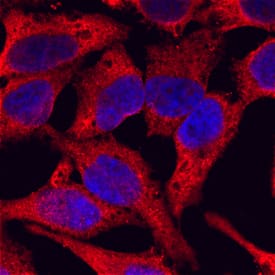
gp96/HSP90B1 in HeLa Human Cell Line. gp96/HSP90B1 was detected in immersion fixed HeLa human cervical epithelial carcinoma cell line using Mouse Anti-Human gp96/HSP90B1 Monoclonal Antibody (Catalog # MAB7606) at 10 µg/mL for 3 hours at room temperature. Cells were stained using the NorthernLights™ 557-conjugated Anti-Mouse IgG Secondary Antibody (red; Catalog # NL007) and counterstained with DAPI (blue). Specific staining was localized to cytoplasm. View our protocol for Fluorescent ICC Staining of Cells on Coverslips.
 View Larger
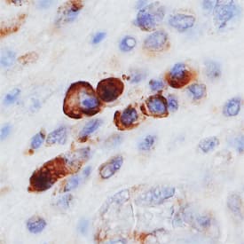
gp96/HSP90B1 in Human Mesothelioma. gp96/HSP90B1 was detected in immersion fixed paraffin-embedded sections of human mesothelioma using Mouse Anti-Human gp96/HSP90B1 Monoclonal Antibody (Catalog # MAB7606) at 15 µg/mL overnight at 4 °C. Before incubation with the primary antibody, tissue was subjected to heat-induced epitope retrieval using Antigen Retrieval Reagent-Basic (Catalog # CTS013). Tissue was stained using the Anti-Mouse HRP-DAB Cell & Tissue Staining Kit (brown; Catalog # CTS002) and counterstained with hematoxylin (blue). Specific staining was localized to plasma membranes and cytoplasm. View our protocol for Chromogenic IHC Staining of Paraffin-embedded Tissue Sections.
 View Larger
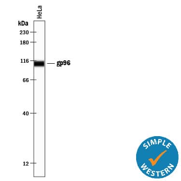
Detection of Human gp96/HSP90B1 by Simple WesternTM. Simple Western lane view shows lysates of HeLa human cervical epithelial carcinoma cell line, loaded at 0.5 mg/mL. A specific band was detected for gp96/HSP90B1 at approximately 108 kDa (as indicated) using 2 µg/mL of Mouse Anti-Human gp96/HSP90B1 Monoclonal Antibody (Catalog # MAB7606). This experiment was conducted under reducing conditions and using the 12-230 kDa separation system.
 View Larger
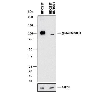
Western Blot Shows Human gp96/HSP90B1 Specificity by Using Knockout Cell Line. Western blot shows lysates of HEK293T human embryonic kidney parental cell line and gp96/HSP90B1 knockout HEK293T cell line (KO). PVDF membrane was probed with 0.2 µg/mL of Mouse Anti-Human gp96/HSP90B1 Monoclonal Antibody (Catalog # MAB7606) followed by HRP-conjugated Anti-Mouse IgG Secondary Antibody (Catalog # HAF018). A specific band was detected for gp96/HSP90B1 at approximately 100 kDa (as indicated) in the parental HEK293T cell line, but is not detectable in knockout HEK293T cell line. GAPDH (Catalog # MAB5718) is shown as a loading control. This experiment was conducted under reducing conditions and using Immunoblot Buffer Group 1.
Reconstitution Calculator
Preparation and Storage
- 12 months from date of receipt, -20 to -70 °C as supplied.
- 1 month, 2 to 8 °C under sterile conditions after reconstitution.
- 6 months, -20 to -70 °C under sterile conditions after reconstitution.
Background: gp96/HSP90B1
Glycoprotein 96 (gp96; also endoplasmin, GRP-94, TRA1 and HSP90B1) is a 94-100 kDa member of the HSP 90 family of proteins. gp96 is a ubiquitously-expressed ER resident protein that is found in a preformed complex with BiP, CaBP1 and UDP-glucosyltransferase. This is a chaperone complex that binds unfolded protein substrates. When folded properly, the substrate is forwarded to calnexin-containing chaperone complexes that promote its maturation. gp96 clients are restricted and include disulfide-bonded integrins, TLRs, LDLR and CD180. Within the complex, gp96 exists as a disulfide-linked homodimer that may form higher-order oligomers. gp96 also appears on the cell surface and may serve as a receptor for bacteria. Mature human gp96 is a 782 amino acid (aa) membrane-associated protein
(aa 22-803). It is not a transmembrane protein but utilizes an ER retention signal (aa 800-803) to interact with the ER membrane. The molecule possesses a
HATPase-C like region (aa 98-219) plus multiple ATP binding and two utilized phosphorylation sites. Over aa 503-660, human gp96 shares 98% aa sequence identity with mouse gp96.
Product Datasheets
Citations for Human gp96/HSP90B1 Antibody
R&D Systems personnel manually curate a database that contains references using R&D Systems products. The data collected includes not only links to publications in PubMed, but also provides information about sample types, species, and experimental conditions.
4
Citations: Showing 1 - 4
Filter your results:
Filter by:
-
Quantitative glycoproteomics reveals new classes of STT3A- and STT3B-dependent N-glycosylation sites
Authors: Natalia A. Cherepanova, Sergey V. Venev, John D. Leszyk, Scott A. Shaffer, Reid Gilmore
Journal of Cell Biology
-
Identification of RNA Content of CHO-derived Extracellular Vesicles from a Production Process
Authors: DJ Busch, Y Zhang, A Kumar, SC Huhn, Z Du, R Liu
Journal of biotechnology, 2022-03-12;0(0):.
Species: Hamster, Virus
Sample Types: Cell Lysates
Applications: Western Blot -
Noise Exposures Causing Hearing Loss Generate Proteotoxic Stress and Activate the Proteostasis Network
Authors: N Jongkamonw, MA Ramirez, S Edassery, ACY Wong, J Yu, T Abbott, K Pak, AF Ryan, JN Savas
Cell Rep, 2020-11-24;33(8):108431.
Species: Mouse
Sample Types: Whole Tissue
Applications: IHC-Fr -
Microdomains form on the luminal face of neuronal extracellular vesicle membranes
Authors: D Matthies, NYJ Lee, I Gatera, HA Pasolli, X Zhao, H Liu, D Walpita, Z Liu, Z Yu, MS Ioannou
Sci Rep, 2020-07-20;10(1):11953.
Species: Rat
Sample Types: Cell Culture Lysates
Applications: Western Blot
FAQs
No product specific FAQs exist for this product, however you may
View all Antibody FAQsReviews for Human gp96/HSP90B1 Antibody
Average Rating: 5 (Based on 2 Reviews)
Have you used Human gp96/HSP90B1 Antibody?
Submit a review and receive an Amazon gift card.
$25/€18/£15/$25CAN/¥75 Yuan/¥2500 Yen for a review with an image
$10/€7/£6/$10 CAD/¥70 Yuan/¥1110 Yen for a review without an image
Filter by:
W-B of RM1 cell lysate
