Human/Mouse/Rat Galectin-3 Antibody Summary
Ala2-Ile264
Accession # P16110
Applications
Mouse Galectin-3 Sandwich Immunoassay
Please Note: Optimal dilutions should be determined by each laboratory for each application. General Protocols are available in the Technical Information section on our website.
Scientific Data
 View Larger
Detection of Mouse Galectin‑3 by Western Blot. Western blot shows lysates of 4T1 mouse breast cancer cell line, Balb/3T3 mouse embryonic fibroblast cell line and HT-2 mouse T cell line. PVDF membrane was probed with 0.25 µg/mL of Rat Anti-Mouse Galectin-3 Monoclonal Antibody (Catalog # MAB1197) followed by HRP-conjugated Anti-Rat IgG Secondary Antibody (Catalog # HAF005). A specific band was detected for Galectin-3 at approximately 28 kDa (as indicated). This experiment was conducted under reducing conditions and using Immunoblot Buffer Group 1.
 View Larger
Detection of Human and Rat Galectin‑3 by Western Blot. Western blot shows lysates of MCF-7 human breast cancer cell line, U-118-MG human glioblastoma/astrocytoma cell line, C6 rat glioma cell line, and NR8383 rat alveolar macrophage cell line. PVDF membrane was probed with 0.5 µg/mL of Rat Anti-Mouse Galectin-3 Monoclonal Antibody (Catalog # MAB1197) followed by HRP-conjugated Anti-Rat IgG Secondary Antibody (Catalog # HAF005). A specific band was detected for Galectin-3 at approximately 28 kDa (as indicated). This experiment was conducted under reducing conditions and using Immunoblot Buffer Group 1.
 View Larger
Detection of Mouse Galectin‑3 by Simple WesternTM. Simple Western lane view shows lysates of HT-2 mouse T cell line and Balb/3T3 mouse embryonic fibroblast cell line, loaded at 0.2 mg/mL. A specific band was detected for Galectin-3 at approximately 43 kDa (as indicated) using 10 µg/mL of Rat Anti-Mouse Galectin-3 Monoclonal Antibody (Catalog # MAB1197) followed by 1:50 dilution of HRP-conjugated Anti-Rat IgG Secondary Antibody (Catalog # HAF005). This experiment was conducted under reducing conditions and using the 12-230 kDa separation system.
 View Larger
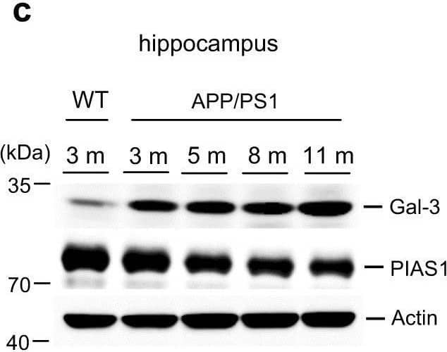
Detection of Galectin-3 by Western Blot A beta oligomerization and Gal-3 expression are age-dependently increased in APP/PS1 mice. a Wild-type (3 months old) and APP/PS1 mice of different ages (3, 5, 8, and 11 months) were sacrificed and dissected hippocampal tissues were subjected to Western blot analysis for endogenous A beta oligomers. b Quantified results for HMW and LMW A beta oligomerization [n = 4 per group; F(4,15) = 39.3, P < 0.001 for HMW and F(4,15) = 74.07, P < 0.001 for LMW]. Statistical significances for various comparisons are shown in the figure. c Western blot analysis showing the expression levels of Gal-3 and PIAS1 in hippocampal samples from the same batch of WT mice and APP/PS1 mice of different ages (n = 4 per group). d Quantified results for Gal-3 expression in WT and APP/PS1 mice [F(4,15) = 14.59, P < 0.001]. e Quantified results for PIAS1 expression in WT and APP/PS1 mice [F(4,15) = 5.86, P < 0.01] The letter “m” indicates month. Statistical significances of various comparisons are shown in the figure. Data are expressed as mean ± SEM. *P < 0.05, **P < 0.01, #P < 0.001 Image collected and cropped by CiteAb from the following open publication (https://pubmed.ncbi.nlm.nih.gov/31127200), licensed under a CC-BY license. Not internally tested by R&D Systems.
 View Larger
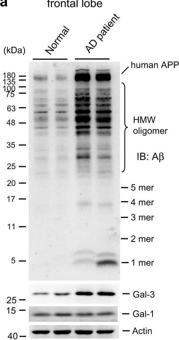
Detection of Galectin-3 by Western Blot A beta oligomerization and Gal-3 expression are increased in AD patients. The frontal lobe lysates of normal subjects and AD patients were subjected to Western blot analysis for A beta oligomerization and the expression of Gal-3 and Gal-1 (n = 4 per group). b–d Quantified results for (b) HMW [t(1,6) = 9.72, P < 0.001] and LMW [t(1,6) = 7.21, P < 0.001] A beta oligomerization, c Gal-3 expression [t(1,6) = 4.92, P < 0.01] and d Gal-1 expression [t(1,6) = 0.43, P > 0.05]. e Immunohistochemical analysis showing the distributions of Gal-3 and ProteoStat staining in normal subjects and AD patients (n = 3 per group). Scale bar, 25 μm. DAPI was used to stain nuclei. Data are expressed as mean ± SEM. **P < 0.01, #P < 0.001 Image collected and cropped by CiteAb from the following open publication (https://pubmed.ncbi.nlm.nih.gov/31127200), licensed under a CC-BY license. Not internally tested by R&D Systems.
 View Larger
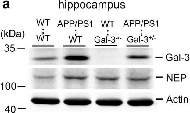
Detection of Galectin-3 by Western Blot Endogenous Gal-3 expression is decreased but memory performance is improved in APP/PS1;Gal-3+/− mice. a Endogenous expression levels of Gal-3 and NEP were examined by Western blot analysis in 3-month-old WT;WT, APP/PS1;WT, WT;Gal-3−/− and APP/PS1;Gal-3+/− mice. b Quantified results for Gal-3 and NEP expression [n = 4 per group; for Gal-3, F(3,12) = 53.47, P < 0.001; q = 10.43, P < 0.001 for the WT;WT group versus the APP/PS1;WT group; q = 6.46, P < 0.001 for the APP/PS1;WT group versus the APP/PS1;Gal-3+/− group and q = 3.98, P < 0.05 for the WT;WT group versus the APP/PS1;Gal-3+/− group; for NEP, F(3,12) = 27.76, P < 0.001; q = 12.04, P < 0.001 for the WT;WT group versus the APP/PS1;WT group; q = 9.72, P < 0.001 for the WT;WT group versus the WT;Gal-3−/− group and q = 6.29, P < 0.01 for the WT;WT group versus the APP/PS1;Gal-3+/− group]. c Acquisition performance obtained by assessing the water maze learning of 8-month-old mice of the four genotypes [n = 7 per group; F(3,24) = 28.72, P < 0.001; q = 9.33, P < 0.001 for the APP/PS1;WT group versus the WT;WT group and q = 3.59, P < 0.05 for the APP/PS1;Gal-3+/− group versus the APP/PS1;WT group]. d Retention performance of the same mice described in (c) [n = 7 per group; F(3,24) = 4.76, P < 0.01; q = 3.94, P < 0.05 for the APP/PS1;WT group versus the WT;WT group; q = 3.25, P < 0.05 for the APP/PS1;Gal-3+/− group versus the APP/PS1;WT group for the target quadrant]. Representative swim patterns from each group are also shown (upper panel). Data are expressed as mean ± SEM. *P < 0.05, **P < 0.01, #P < 0.001 Image collected and cropped by CiteAb from the following open publication (https://pubmed.ncbi.nlm.nih.gov/31127200), licensed under a CC-BY license. Not internally tested by R&D Systems.
 View Larger
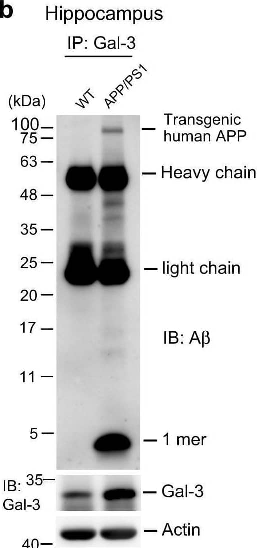
Detection of Galectin-3 by Western Blot Galectin-3 associates with A beta and interacts with A beta. a Immunohistochemical analysis showing the distributions of endogenous Gal-3 and A beta in hippocampal samples of WT (left panel) and APP/PS1 (right panel) mice at 11 months of age. Scale bar, 200 μm. b Co-IP experiment showing preferential association of Gal-3 with endogenous A beta monomers in 8-month-old APP/PS1 mice. Experiments were performed in duplicate. c Different amounts of recombinant human Gal-3 protein (0.25, 0.5, 1, and 2 μg) were added to a solution containing A beta 42 peptide (15 μg), the mixture was subjected to a thioflavin-T assay, and fluorescence was measured hourly at different time points. The fluorescence plateaued at the 7-h time point. Experiments were performed in triplicate. The abbreviation “rh” indicates “recombinant human”. Data are expressed as mean ± SEM Image collected and cropped by CiteAb from the following open publication (https://pubmed.ncbi.nlm.nih.gov/31127200), licensed under a CC-BY license. Not internally tested by R&D Systems.
 View Larger
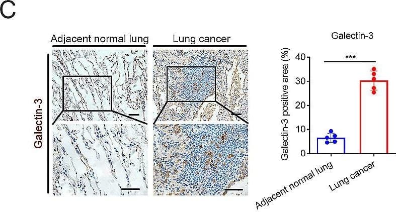
Detection of Galectin-3 by Immunohistochemistry Tumor derived galectin-3 is a ligand for TREM2. (C) Immunohistochemical staining of galectin-3 in adjacent normal lung & intratumor areas of lung tissues (n = 5). Scale bars, 20 μm. Image collected & cropped by CiteAb from the following open publication (https://pubmed.ncbi.nlm.nih.gov/39135069), licensed under a CC-BY license. Not internally tested by R&D Systems.
Reconstitution Calculator
Preparation and Storage
- 12 months from date of receipt, -20 to -70 °C as supplied.
- 1 month, 2 to 8 °C under sterile conditions after reconstitution.
- 6 months, -20 to -70 °C under sterile conditions after reconstitution.
Background: Galectin-3
The galectins constitute a large family of carbohydrate-binding proteins with specificity for N-acetyl-lactosamine-containing glycoproteins. At least 14 mammalian galectins, which share structural similarities in their carbohydrate recognition domains (CRD), have been identified. The galectins have been classified into the prototype galectins (-1, -2, -5, -7, -10, -11, -13, -14), which contain one CRD and exist either as a monomer or a noncovalent homodimer; the chimera galectins (Galectin-3) containing one CRD linked to a nonlectin domain; and the tandem-repeat galectins (-4, -6, -8, -9, -12) consisting of two CRDs joined by a linker peptide. Galectins lack a classical signal peptide and can be localized to the cytosolic compartments where they have intracellular functions. However, via one or more as yet unidentified non-classical secretory pathways, galectins can also be secreted to function extracellularly. Individual members of the galectin family have different tissue distribution profiles and exhibit subtle differences in their carbohydrate-binding specificities. Each family member may preferentially bind to a unique subset of cell-surface glycoproteins (1-4). Galectin-3, also known as Mac-2, L29, CBP35, and epsilon BP, is a chimera galectin that has a tendency to dimerize. Besides the soluble protein, alternatively spliced forms of chicken Galectin-3 containing a transmembrane-spanning domain and a leucine zipper motif have been reported. Galectin-3 is expressed in tumor cells, macrophages, activated T cells, osteoclasts, epithelial cells, and fibroblasts. It binds various matrix glycoproteins including laminin, fibronectin, LAMPS, 90K/Mac-2BP, MP20, and CEA. Galectin-3 promotes cell growth and proliferation for many cell types. Galectin-3 acts intracellularly to prevent apoptosis. Depending on the cell types, Galectin-3 exhibits pro- or anti-adhesive properties. Galectin-3 has proinflammatory activities in vitro and in vivo. It induces pro-inflammatory and inhibits Th2 type cytokine production. Galectin-3 chemoattracts monocytes and macrophages. It activates and degranulates basophils and mast cells. Elevated circulating levels of Galectin-3 has been show to correlate with the malignant potential of several types of cancer, suggesting that Galectin-3 is also involved in tumor growth and metastasis. Human and mouse Galectin-3 shares approximately 80% amino acid sequence similarity (1-5).
- Rabinovich, A. et al. (2002) Trends in Immunol. 23:313.
- Rabinovich, A. et al. (2002) J. Leukocyte Biology 71:741.
- Hughes, R.C. (2001) Biochimie 83:667.
- R&D Systems Cytokine Bulletin; Summer 2002.
- Gorski, J.P. et al. (2002) J. Biol. Chem. 277:18840.
Product Datasheets
Citations for Human/Mouse/Rat Galectin-3 Antibody
R&D Systems personnel manually curate a database that contains references using R&D Systems products. The data collected includes not only links to publications in PubMed, but also provides information about sample types, species, and experimental conditions.
6
Citations: Showing 1 - 6
Filter your results:
Filter by:
-
Galectin-3 Negatively Regulates Hippocampus-Dependent Memory Formation through Inhibition of Integrin Signaling and Galectin-3 Phosphorylation
Authors: Yan-Chu Chen, Yun-Li Ma, Cheng-Hsiung Lin, Sin-Jhong Cheng, Wei-Lun Hsu, Eminy H.-Y. Lee
Frontiers in Molecular Neuroscience
-
Postpartum administration of eplerenone to mitigate vascular dysfunction in mice following a preeclampsia-like pregnancy
Authors: Binder, NK;de Alwis, N;Beard, S;Fato, BR;Garg, A;Baird, L;Young, MJ;Hannan, NJ;
Scientific reports
Species: Mouse
Sample Types: Whole Tissue
Applications: Immunohistochemistry -
Galectin-3 promotes A&beta oligomerization and A&beta toxicity in a mouse model of Alzheimer's disease
Authors: CC Tao, KM Cheng, YL Ma, WL Hsu, YC Chen, JL Fuh, WJ Lee, CC Chao, EHY Lee
Cell Death Differ., 2019-05-24;27(1):192-209.
Species: Rat
Sample Types: Hippocampal Cell Lysates
Applications: Western Blot -
Proteomic and functional analysis identifies galectin-1 as a novel regulatory component of the cytotoxic granule machinery
Authors: T Clemente, NJ Vieira, JP Cerliani, C Adrain, A Luthi, MR Dominguez, M Yon, FC Barrence, TB Riul, RD Cummings, TM Zorn, S Amigorena, M Dias-Baruf, MM Rodrigues, SJ Martin, GA Rabinovich, GP Amarante-M
Cell Death Dis, 2017-12-07;8(12):e3176.
Species: Human
Sample Types: Cell Lysates
Applications: Western Blot -
Galectin-3 negatively regulates the frequency and function of CD4(+) CD25(+) Foxp3(+) regulatory T cells and influences the course of Leishmania major infection.
Authors: Fermino M, Dias F, Lopes C, Souza M, Cruz A, Liu F, Chammas R, Roque-Barreira M, Rabinovich G, Bernardes E
Eur J Immunol, 2013-05-17;43(7):1806-17.
Species: Mouse
Sample Types: Whole Tissue
Applications: IHC-Fr -
The receptor of advanced glycation end products plays a central role in advanced oxidation protein products-induced podocyte apoptosis.
Kidney Int, 2012-05-23;82(7):759-70.
Species: Mouse
Sample Types: Whole Cells
Applications: Neutralization
FAQs
No product specific FAQs exist for this product, however you may
View all Antibody FAQsReviews for Human/Mouse/Rat Galectin-3 Antibody
There are currently no reviews for this product. Be the first to review Human/Mouse/Rat Galectin-3 Antibody and earn rewards!
Have you used Human/Mouse/Rat Galectin-3 Antibody?
Submit a review and receive an Amazon gift card.
$25/€18/£15/$25CAN/¥75 Yuan/¥2500 Yen for a review with an image
$10/€7/£6/$10 CAD/¥70 Yuan/¥1110 Yen for a review without an image
