
Key Product Details
Species Reactivity
Validated:
Human, Mouse, Rat
Cited:
Human, Mouse, Rat, Porcine
Applications
Validated:
Immunohistochemistry, Western Blot, Immunocytochemistry, Simple Western
Cited:
Western Blot, Immunocytochemistry, Co-Immunoprecipitation, Simple Plex Automated Immunoassay
Label
Unconjugated
Antibody Source
Monoclonal Mouse IgG1 Clone # 686613
Loading...
Product Specifications
Immunogen
E. coli-derived recombinant human GAPDH/G3PDH
Met1-Ala150
Accession # P04406
Met1-Ala150
Accession # P04406
Specificity
Detects human, mouse and rat GAPDH/G3PDH in Western blots. In direct ELISAs, 100% cross-reactivity with recombinant mouse GAPDH/G3PDH and no cross-reactivity with recombinant human GAPDH-2 is observed.
Clonality
Monoclonal
Host
Mouse
Isotype
IgG1
Scientific Data Images for Human/Mouse/Rat GAPDH Antibody
Detection of Human, Mouse, and Rat GAPDH/G3PDH by Western Blot.
Western blot shows lysates of human brain tissue, mouse brain tissue, and rat brain tissue. PVDF Membrane was probed with 0.05 µg/mL of Mouse Anti-Human/Mouse/Rat GAPDH/G3PDH Monoclonal Antibody (Catalog # MAB5718) followed by HRP-conjugated Anti-Mouse IgG Secondary Antibody (Catalog # HAF007). A specific band was detected for GAPDH/G3PDH at approximately 39 kDa (as indicated). This experiment was conducted under reducing conditions and using Immunoblot Buffer Group 2.
GAPDH in HeLa Human Cell Line.
GAPDH was detected in immersion fixed HeLa human cervical epithelial carcinoma cell line using Mouse Anti-Human/Mouse/Rat GAPDH Monoclonal Antibody (Catalog # MAB5718) at 8 µg/mL for 3 hours at room temperature. Cells were stained using the NorthernLights™ 557-conjugated Anti-Mouse IgG Secondary Antibody (red; Catalog # NL007) and counterstained with DAPI (blue). View our protocol for Fluorescent ICC Staining of Cells on Coverslips.
GAPDH/G3PDH in Human Kidney.
GAPDH/G3PDH was detected in immersion fixed paraffin-embedded sections of human kidney using Mouse Anti-Human/Mouse/Rat GAPDH/G3PDH Monoclonal Antibody (Catalog # MAB5718) at 15 µg/mL overnight at 4 °C. Before incubation with the primary antibody, tissue was subjected to heat-induced epitope retrieval using Antigen Retrieval Reagent-Basic (Catalog # CTS013). Tissue was stained using the Anti-Mouse HRP-DAB Cell & Tissue Staining Kit (brown; Catalog # CTS002) and counterstained with hematoxylin (blue). Specific staining was localized to cytoplasm and nuclei. View our protocol for Chromogenic IHC Staining of Paraffin-embedded Tissue Sections.
Detection of Human and Mouse GAPDH/G3PDH by Simple WesternTM.
Simple Western lane view shows lysates of Jurkat human acute T cell leukemia cell line and C2C12 mouse myoblast cell line, loaded at 0.2 mg/mL. A specific band was detected for GAPDH/G3PDH at approximately 41 kDa (as indicated) using 0.25 µg/mL of Mouse Anti-Human/Mouse/Rat GAPDH/G3PDH Monoclonal Antibody (Catalog # MAB5718). This experiment was conducted under reducing conditions and using the 12-230 kDa separation system.
Detection of Human, Mouse, and Rat GAPDH by Simple WesternTM.
Simple Western lane view shows lysates of Jurkat human acute T cell leukemia cell line, HeLa human cervical epithelial carcinoma cell line, C2C12 mouse myoblast cell line, and C6 rat glioma cell line, loaded at 0.2 mg/mL. A specific band was detected for GAPDH at approximately 42 kDa (as indicated) using 1 µg/mL of Mouse Anti-Human/Mouse/Rat GAPDH Monoclonal Antibody (Catalog # MAB5718). This experiment was conducted under reducing conditions and using the 12-230 kDa separation system.
Detection of Human GAPDH by Western Blot
Simultaneous knockdown of multiple genes in primary human islets following electroporation. (A) Graph shows RT-qPCR results of islets electroporated with non-targeting control siRNA or siRNA targeting RB and p130 (L-003299). (B) Graph shows RT-qPCR results of islets electroporated with non-targeted control siRNA or siRNAs targeting the indicated gene products. The housekeeping gene GAPDH was used as the control. Representative experiment shown from 3 separate experiments. Error bars indicate standard deviation. (C) Western blot for RB protein in human islet lysates after electroporation of control or targeting siRNA. GAPDH was used as a loading control. RB appears as a characteristic doublet representing the hypo and hyperphosphorylated protein. Representative experiment shown from 3 different experiments. Image collected and cropped by CiteAb from the following open publication (https://pubmed.ncbi.nlm.nih.gov/25305068), licensed under a CC-BY license. Not internally tested by R&D Systems.
Detection of GAPDH by Western Blot
Evaluation of CPP/mRNA-mediated protein expression. (A) Representative fluorescence images and a quantitative graph showing mCherry-positive CT26.CL25 cells 24 h after treatment with CPP/mCherry mRNA complexes (6.8 nM). Scale bar: 275 μm. Data are presented as the mean ± SD (n = 3). (B) Representative flow cytometry histograms and graphs of EGFP-positive CL26.CL25 cells transfected with CPP/EGFP mRNA complexes (6.8 nM). Data are presented as the mean ± SD (n = 3). (C) Western blot analysis of the expression levels of OVA and GAPDH in CT26.CL25 cells. Image collected and cropped by CiteAb from the following open publication (https://pubmed.ncbi.nlm.nih.gov/35745843), licensed under a CC-BY license. Not internally tested by R&D Systems.
Detection of GAPDH by Western Blot
Molecular effect of intraluminal chloride and GsMTx4 in smooth muscle cells. a-c Upper panel represents the main cumulative effect of intraluminal chloride concentration ([Cl−]) and medium supplementation with and without GsMTx4. a Examples of representative blots are shown. b, c Protein expression levels for b myosin light chain 2 (MLC2), and c alpha-smooth muscle actin ( alpha -SMA) are quantified. d–f Lower panel shows the additional effect of PIEZO1/2 inhibition after intraluminal injection of Cl− channels inhibitors: cystic fibrosis transmembrane conductance regulator inhibitor172 (CFTRinh) to CFTR and; calcium-dependent Cl− channel inhibitor A01 (CaCCinh) to CaCCs. d Examples of representative blots are shown. e–f Relative expression levels of e MLC2, and f alpha -SMA are displayed. 143 mM Cl− and standard solution (SS) represent the control condition for [Cl−] and Cl− channels inhibitors, respectively. White and dotted rectangles represent the medium supplementation with and without GsMTx4, respectively. Each lane represents a pooled-tissue sample, and the relative expression levels were determined against GAPDH. n ≥ 4 were used per antibody/condition. Results are presented as mean ± SD. Symbols indicate the main effects and non-redundant interactions of the two-way ANOVA. p < alpha 0.0001, beta 0.001, gamma 0.01, µ0.05 Image collected and cropped by CiteAb from the following open publication (https://pubmed.ncbi.nlm.nih.gov/36740669), licensed under a CC-BY license. Not internally tested by R&D Systems.
Detection of GAPDH by Western Blot
The impact of combination therapy on protein and gene expression in SJSA-1 osteosarcoma cells. (A) A Western blot analysis illustrating the effects of the combination treatment on key proteins involved in metastasis, cell survival, and apoptosis in SJSA-1 osteosarcoma cells. The expression levels of MMP9 and MMP13, which facilitate tumor cell invasion and metastasis through ECM degradation, were significantly reduced, indicating a potential inhibitory effect of the combination therapy on metastatic progression. Additionally, phosphorylated AKT (pAKT), a central regulator of the PI3K/AKT pathway associated with cell survival and chemoresistance, and runt-related transcription factor 2 (RUNX2), a critical modulator of osteosarcoma progression and tumor aggressiveness, were both downregulated, suggesting the suppression of key oncogenic pathways. Conversely, apoptotic markers, including cleaved caspase-8, cleaved caspase-3, and total caspase-3, exhibited increased activation following treatment, indicating enhanced apoptotic signaling. GAPDH was used as a loading control to ensure equal protein loading across samples. (B) A densitometric analysis of the Western blot bands representing the expression levels of target proteins after 48 h of treatment. The band intensities were quantified using Image Lab software (Version 6.1.0 build 7) and normalized to the GAPDH. Relative protein expression levels are expressed as a fold change compared to the untreated control group. The data represent the mean ± the standard deviation from two independent experiments. (C) qRT-PCR analyzed the gene expression of the apoptosis-related markers p16 and p53 following treatment. The results highlight the upregulation of p16 and stable expression of p53 in treated cells. (* p < 0.05 and ** p < 0.01). Image collected and cropped by CiteAb from the following open publication (https://pubmed.ncbi.nlm.nih.gov/40332124), licensed under a CC-BY license. Not internally tested by R&D Systems.Applications for Human/Mouse/Rat GAPDH Antibody
Application
Recommended Usage
Immunocytochemistry
8-25 µg/mL
Sample: Immersion fixed HeLa human cervical epithelial carcinoma cell line
Sample: Immersion fixed HeLa human cervical epithelial carcinoma cell line
Immunohistochemistry
8-25 µg/mL
Sample: Immersion fixed paraffin-embedded sections of human kidney
Sample: Immersion fixed paraffin-embedded sections of human kidney
Simple Western
0.25 - 1 µg/mL
Sample: Jurkat human acute T cell leukemia cell line, HeLa human cervical epithelial carcinoma cell line, C2C12 mouse myoblast cell line, and C6 rat glioma cell line
Sample: Jurkat human acute T cell leukemia cell line, HeLa human cervical epithelial carcinoma cell line, C2C12 mouse myoblast cell line, and C6 rat glioma cell line
Western Blot
0.05 µg/mL
Sample: Human brain tissue, mouse brain tissue, and rat brain tissue
Sample: Human brain tissue, mouse brain tissue, and rat brain tissue
Reviewed Applications
Read 4 reviews rated 5 using MAB5718 in the following applications:
Formulation, Preparation, and Storage
Purification
Protein A or G purified from hybridoma culture supernatant
Reconstitution
Sterile PBS to a final concentration of 0.5 mg/mL. For liquid material, refer to CoA for concentration.
Loading...
Formulation
Lyophilized from a 0.2 μm filtered solution in PBS with Trehalose. *Small pack size (SP) is supplied either lyophilized or as a 0.2 µm filtered solution in PBS.
Shipping
Lyophilized product is shipped at ambient temperature. Liquid small pack size (-SP) is shipped with polar packs. Upon receipt, store immediately at the temperature recommended below.
Stability & Storage
Use a manual defrost freezer and avoid repeated freeze-thaw cycles.
- 12 months from date of receipt, -20 to -70 °C as supplied.
- 1 month, 2 to 8 °C under sterile conditions after reconstitution.
- 6 months, -20 to -70 °C under sterile conditions after reconstitution.
Calculators
Background: GAPDH
aa 1‑150, human GAPDH shares 92% aa identity with mouse GAPDH.
Long Name
Glyceraldehyde-3-phosphate Dehydrogenase
Alternate Names
G3PDH
Gene Symbol
GAPDH
UniProt
Additional GAPDH Products
Product Documents for Human/Mouse/Rat GAPDH Antibody
Certificate of Analysis
To download a Certificate of Analysis, please enter a lot or batch number in the search box below.
Note: Certificate of Analysis not available for kit components.
Product Specific Notices for Human/Mouse/Rat GAPDH Antibody
For research use only
Related Research Areas
Citations for Human/Mouse/Rat GAPDH Antibody
 Powered by Bioz
Customer Reviews for Human/Mouse/Rat GAPDH Antibody (4)
5 out of 5
4 Customer Ratings
Have you used Human/Mouse/Rat GAPDH Antibody?
Submit a review and receive an Amazon gift card!
$25/€18/£15/$25CAN/¥2500 Yen for a review with an image
$10/€7/£6/$10CAN/¥1110 Yen for a review without an image
Submit a review
Customer Images
Showing
1
-
4 of
4 reviews
Showing All
Filter By:
-
Application: Western BlotSample Tested: Cell LinesSpecies: HumanVerified Customer | Posted 08/05/2025

-
Application: Western BlotSample Tested: Cancer cell lysates and Xenograft LysatesSpecies: Human and MouseVerified Customer | Posted 07/03/2018
-
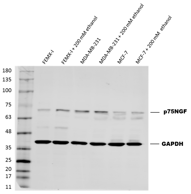
Application: Western BlotSample Tested: Human cell lineSpecies: HumanVerified Customer | Posted 03/01/2017Expression of Human p75NGF in several cell lines, with GAPDH levels showing equal loading. Dilution: 1:10,000 in PBS with 5% BSA. Secondary Ab: 1:5,000 anti-mouse IgG.
-
Application: Western BlotSample Tested: See PMID 24040198Species: MouseVerified Customer | Posted 02/25/2015
There are no reviews that match your criteria.
Protocols
Find general support by application which include: protocols, troubleshooting, illustrated assays, videos and webinars.
- Antigen Retrieval Protocol (PIER)
- Antigen Retrieval for Frozen Sections Protocol
- Appropriate Fixation of IHC/ICC Samples
- Cellular Response to Hypoxia Protocols
- Chromogenic IHC Staining of Formalin-Fixed Paraffin-Embedded (FFPE) Tissue Protocol
- Chromogenic Immunohistochemistry Staining of Frozen Tissue
- ClariTSA™ Fluorophore Kits
- Detection & Visualization of Antibody Binding
- Fluorescent IHC Staining of Frozen Tissue Protocol
- Graphic Protocol for Heat-induced Epitope Retrieval
- Graphic Protocol for the Preparation and Fluorescent IHC Staining of Frozen Tissue Sections
- Graphic Protocol for the Preparation and Fluorescent IHC Staining of Paraffin-embedded Tissue Sections
- Graphic Protocol for the Preparation of Gelatin-coated Slides for Histological Tissue Sections
- ICC Cell Smear Protocol for Suspension Cells
- ICC Immunocytochemistry Protocol Videos
- ICC for Adherent Cells
- IHC Sample Preparation (Frozen sections vs Paraffin)
- Immunocytochemistry (ICC) Protocol
- Immunocytochemistry Troubleshooting
- Immunofluorescence of Organoids Embedded in Cultrex Basement Membrane Extract
- Immunofluorescent IHC Staining of Formalin-Fixed Paraffin-Embedded (FFPE) Tissue Protocol
- Immunohistochemistry (IHC) and Immunocytochemistry (ICC) Protocols
- Immunohistochemistry Frozen Troubleshooting
- Immunohistochemistry Paraffin Troubleshooting
- Preparing Samples for IHC/ICC Experiments
- Preventing Non-Specific Staining (Non-Specific Binding)
- Primary Antibody Selection & Optimization
- Protocol for Heat-Induced Epitope Retrieval (HIER)
- Protocol for Making a 4% Formaldehyde Solution in PBS
- Protocol for VisUCyte™ HRP Polymer Detection Reagent
- Protocol for the Fluorescent ICC Staining of Cell Smears - Graphic
- Protocol for the Fluorescent ICC Staining of Cultured Cells on Coverslips - Graphic
- Protocol for the Preparation & Fixation of Cells on Coverslips
- Protocol for the Preparation and Chromogenic IHC Staining of Frozen Tissue Sections
- Protocol for the Preparation and Chromogenic IHC Staining of Frozen Tissue Sections - Graphic
- Protocol for the Preparation and Chromogenic IHC Staining of Paraffin-embedded Tissue Sections
- Protocol for the Preparation and Chromogenic IHC Staining of Paraffin-embedded Tissue Sections - Graphic
- Protocol for the Preparation and Fluorescent ICC Staining of Cells on Coverslips
- Protocol for the Preparation and Fluorescent ICC Staining of Non-adherent Cells
- Protocol for the Preparation and Fluorescent ICC Staining of Stem Cells on Coverslips
- Protocol for the Preparation and Fluorescent IHC Staining of Frozen Tissue Sections
- Protocol for the Preparation and Fluorescent IHC Staining of Paraffin-embedded Tissue Sections
- Protocol for the Preparation of Gelatin-coated Slides for Histological Tissue Sections
- Protocol for the Preparation of a Cell Smear for Non-adherent Cell ICC - Graphic
- R&D Systems Quality Control Western Blot Protocol
- TUNEL and Active Caspase-3 Detection by IHC/ICC Protocol
- The Importance of IHC/ICC Controls
- Troubleshooting Guide: Immunohistochemistry
- Troubleshooting Guide: Western Blot Figures
- Western Blot Conditions
- Western Blot Protocol
- Western Blot Protocol for Cell Lysates
- Western Blot Troubleshooting
- Western Blot Troubleshooting Guide
- View all Protocols, Troubleshooting, Illustrated assays and Webinars
Loading...