Human/Mouse/Rat Gelsolin/GSN Antibody
R&D Systems | Catalog # MAB8170

Key Product Details
Validated by
Knockout/Knockdown
Species Reactivity
Validated:
Human, Mouse, Rat
Cited:
Human
Applications
Validated:
Knockout Validated, Multiplex Immunofluorescence, Immunohistochemistry, Western Blot, Simple Western, COMET
Cited:
Neutralization
Label
Unconjugated
Antibody Source
Monoclonal Mouse IgG1 Clone # 893205
Loading...
Product Specifications
Immunogen
HEK293 human embryonic kidney cell line transfected with human Gelsolin/GSN
Met1-Ala782
Accession # P06396
Met1-Ala782
Accession # P06396
Specificity
Detects human Gelsolin/GSN in ELISAs. Detects human, mouse and rat Gelsolin/GSN in Western Blots.
Clonality
Monoclonal
Host
Mouse
Isotype
IgG1
Scientific Data Images for Human/Mouse/Rat Gelsolin/GSN Antibody
Detection of Gelsolin/GSN in Human Renal Cell Carcinoma via seqIF™ staining on COMET™
Gelsolin/GSN was detected in immersion fixed paraffin-embedded sections of human Renal Cell Carcinoma using Mouse Anti-Human Gelsolin/GSN, pan Monoclonal Antibody (Catalog # MAB8170) at 20ug/mL at 37 ° Celsius for 4 minutes. Before incubation with the primary antibody, tissue underwent an all-in-one dewaxing and antigen retrieval preprocessing using PreTreatment Module (PT Module) and Dewax and HIER Buffer H (pH 9; Epredia Catalog # TA-999-DHBH).Tissue was stained using the Alexa Fluor™ 647 Goat anti-Mouse IgG Secondary Antibody at 1:200 at 37 ° Celsius for 2 minutes. (Yellow; Lunaphore Catalog # DR647MS) and counterstained with DAPI (blue; Lunaphore Catalog # DR100). Specific staining was localized to the cytoplasm. Protocol available in COMET™ Panel Builder.
Detection of Human, Mouse, and Rat Gelsolin/GSN by Western Blot.
Western blot shows lysates of SK-Mel-28 human malignant melanoma cell line, MEF mouse embryonic feeder cells, and NR8383 rat alveolar macrophage cell line. PVDF membrane was probed with 0.5 µg/mL of Mouse Anti-Human/Mouse/Rat Gelsolin/GSN Monoclonal Antibody (Catalog # MAB8170) followed by HRP-conjugated Anti-Mouse IgG Secondary Antibody (Catalog # HAF018). A specific band was detected for Gelsolin/GSN at approximately 95 kDa (as indicated). This experiment was conducted under reducing conditions and using Immunoblot Buffer Group 1.
Detection of Human Gelsolin/GSN by Simple WesternTM.
Simple Western shows lysates of Exosome Standards (HT‑29) (NBP3-11685), Exosome Standards (U‑87 MG) (NBP2-49844) and SK‑Mel‑28 human malignant melanoma cell line, loaded at 0.5 mg/ml. A specific band was detected for Gelsolin/GSN at approximately 90 kDa (as indicated) using 20 µg/mL of Mouse Anti-Human/Mouse/Rat Gelsolin/GSN Monoclonal Antibody (Catalog # MAB8170). This experiment was conducted under reducing conditions and using the 12-230kDa separation system.
Gelsolin/GSN in Human Kidney.
Gelsolin/GSN was detected in formalin fixed paraffin-embedded sections of human kidney using Mouse Anti-Human/Mouse/Rat Gelsolin/GSN Monoclonal Antibody (Catalog # MAB8170) at 15 µg/mL overnight at 4 °C. Tissue was stained using the Anti-Mouse HRP-DAB Cell & Tissue Staining Kit (brown; Catalog # CTS002) and counterstained with hematoxylin (blue). Specific staining was localized to glomeruli and distal convoluted tubules. View our protocol for Chromogenic IHC Staining of Paraffin-embedded Tissue Sections.
Western Blot Shows Human Gelsolin/GSN Specificity Using Knockout Cell Line.
Western blot shows lysates of U2OS human osteosarcoma cell line and Gelsolin/GSN knockout U2OS cell line (KO). Nitrocellulose membrane was probed with 0.1 µg/mL of Mouse Anti-Human/Mouse/Rat Gelsolin/GSN Monoclonal Antibody (Catalog # MAB8170) followed by HRP-conjugated Anti-Mouse IgG Secondary Antibody. A specific band was detected for Gelsolin/GSN at approximately 80 kDa (as indicated) in the parental U2OS cell line, but is not detectable in knockout U2OS cell line. The Ponceau stained transfer of the blot is shown. This experiment was conducted under reducing conditions. Image, protocol, and testing courtesy of YCharOS Inc. See ycharos.com for additional details.Applications for Human/Mouse/Rat Gelsolin/GSN Antibody
Application
Recommended Usage
COMET
Optimal dilutions of this antibody should be experimentally determined.
Immunohistochemistry
8-25 µg/mL
Sample: Formalin fixed paraffin-embedded sections of human kidney
Sample: Formalin fixed paraffin-embedded sections of human kidney
Knockout Validated
Gelsolin/GSN is specifically detected in the parental U2OS cell line, but is not detectable in knockout U2OS cell line.
Multiplex Immunofluorescence
20 µg/mL
Sample: Immersion fixed paraffin-embedded sections of human Renal Cell Carcinoma
Sample: Immersion fixed paraffin-embedded sections of human Renal Cell Carcinoma
Simple Western
5-20 µg/mL
Sample: Exosome Standards (HT-29) (Catalog # NBP3-11685), Exosome Standards (U-87 MG) (Catalog # NBP2-49844) and SK‑Mel‑28 human malignant melanoma cell line
Sample: Exosome Standards (HT-29) (Catalog # NBP3-11685), Exosome Standards (U-87 MG) (Catalog # NBP2-49844) and SK‑Mel‑28 human malignant melanoma cell line
Western Blot
0.5 µg/mL
Sample: SK‑Mel‑28 human malignant melanoma cell line, MEF mouse embryonic feeder cells, and NR8383 rat alveolar macrophage cell line
Sample: SK‑Mel‑28 human malignant melanoma cell line, MEF mouse embryonic feeder cells, and NR8383 rat alveolar macrophage cell line
Reviewed Applications
Read 1 review rated 5 using MAB8170 in the following applications:
Formulation, Preparation, and Storage
Purification
Protein A or G purified from hybridoma culture supernatant
Reconstitution
Reconstitute at 0.5 mg/mL in sterile PBS. For liquid material, refer to CoA for concentration.
Loading...
Formulation
Lyophilized from a 0.2 μm filtered solution in PBS with Trehalose. See Certificate of Analysis for details.
*Small pack size (-SP) is supplied either lyophilized or as a 0.2 µm filtered solution in PBS.
*Small pack size (-SP) is supplied either lyophilized or as a 0.2 µm filtered solution in PBS.
Shipping
Lyophilized product is shipped at ambient temperature. Liquid small pack size (-SP) is shipped with polar packs. Upon receipt, store immediately at the temperature recommended below.
Stability & Storage
Use a manual defrost freezer and avoid repeated freeze-thaw cycles.
- 12 months from date of receipt, -20 to -70 °C as supplied.
- 1 month, 2 to 8 °C under sterile conditions after reconstitution.
- 6 months, -20 to -70 °C under sterile conditions after reconstitution.
Calculators
Background: Gelsolin/GSN
Alternate Names
ADF, AGEL, Brevin, GSN
Gene Symbol
GSN
UniProt
Additional Gelsolin/GSN Products
Product Documents for Human/Mouse/Rat Gelsolin/GSN Antibody
Certificate of Analysis
To download a Certificate of Analysis, please enter a lot or batch number in the search box below.
Note: Certificate of Analysis not available for kit components.
Product Specific Notices for Human/Mouse/Rat Gelsolin/GSN Antibody
For research use only
Related Research Areas
Citations for Human/Mouse/Rat Gelsolin/GSN Antibody
 Powered by Bioz
Customer Reviews for Human/Mouse/Rat Gelsolin/GSN Antibody (1)
5 out of 5
1 Customer Rating
Have you used Human/Mouse/Rat Gelsolin/GSN Antibody?
Submit a review and receive an Amazon gift card!
$25/€18/£15/$25CAN/¥2500 Yen for a review with an image
$10/€7/£6/$10CAN/¥1110 Yen for a review without an image
Submit a review
Customer Images
Showing
1
-
1 of
1 review
Showing All
Filter By:
-
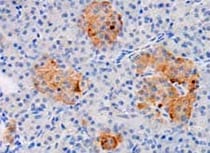
Application: ImmunohistochemistrySample Tested: Pancreas tissueSpecies: MouseVerified Customer | Posted 02/09/2022

There are no reviews that match your criteria.
Protocols
Find general support by application which include: protocols, troubleshooting, illustrated assays, videos and webinars.
- Antigen Retrieval Protocol (PIER)
- Antigen Retrieval for Frozen Sections Protocol
- Appropriate Fixation of IHC/ICC Samples
- Cellular Response to Hypoxia Protocols
- Chromogenic IHC Staining of Formalin-Fixed Paraffin-Embedded (FFPE) Tissue Protocol
- Chromogenic Immunohistochemistry Staining of Frozen Tissue
- ClariTSA™ Fluorophore Kits
- Detection & Visualization of Antibody Binding
- Fluorescent IHC Staining of Frozen Tissue Protocol
- Graphic Protocol for Heat-induced Epitope Retrieval
- Graphic Protocol for the Preparation and Fluorescent IHC Staining of Frozen Tissue Sections
- Graphic Protocol for the Preparation and Fluorescent IHC Staining of Paraffin-embedded Tissue Sections
- Graphic Protocol for the Preparation of Gelatin-coated Slides for Histological Tissue Sections
- IHC Sample Preparation (Frozen sections vs Paraffin)
- Immunofluorescent IHC Staining of Formalin-Fixed Paraffin-Embedded (FFPE) Tissue Protocol
- Immunohistochemistry (IHC) and Immunocytochemistry (ICC) Protocols
- Immunohistochemistry Frozen Troubleshooting
- Immunohistochemistry Paraffin Troubleshooting
- Preparing Samples for IHC/ICC Experiments
- Preventing Non-Specific Staining (Non-Specific Binding)
- Primary Antibody Selection & Optimization
- Protocol for Heat-Induced Epitope Retrieval (HIER)
- Protocol for Making a 4% Formaldehyde Solution in PBS
- Protocol for VisUCyte™ HRP Polymer Detection Reagent
- Protocol for the Preparation & Fixation of Cells on Coverslips
- Protocol for the Preparation and Chromogenic IHC Staining of Frozen Tissue Sections
- Protocol for the Preparation and Chromogenic IHC Staining of Frozen Tissue Sections - Graphic
- Protocol for the Preparation and Chromogenic IHC Staining of Paraffin-embedded Tissue Sections
- Protocol for the Preparation and Chromogenic IHC Staining of Paraffin-embedded Tissue Sections - Graphic
- Protocol for the Preparation and Fluorescent IHC Staining of Frozen Tissue Sections
- Protocol for the Preparation and Fluorescent IHC Staining of Paraffin-embedded Tissue Sections
- Protocol for the Preparation of Gelatin-coated Slides for Histological Tissue Sections
- R&D Systems Quality Control Western Blot Protocol
- TUNEL and Active Caspase-3 Detection by IHC/ICC Protocol
- The Importance of IHC/ICC Controls
- Troubleshooting Guide: Immunohistochemistry
- Troubleshooting Guide: Western Blot Figures
- Western Blot Conditions
- Western Blot Protocol
- Western Blot Protocol for Cell Lysates
- Western Blot Troubleshooting
- Western Blot Troubleshooting Guide
- View all Protocols, Troubleshooting, Illustrated assays and Webinars
Loading...