Human Prolactin Antibody Summary
Leu29-Cys227
Accession # Q5THQ0
*Small pack size (-SP) is supplied either lyophilized or as a 0.2 µm filtered solution in PBS.
Customers also Viewed
Applications
Human Prolactin Sandwich Immunoassay
Please Note: Optimal dilutions should be determined by each laboratory for each application. General Protocols are available in the Technical Information section on our website.
Scientific Data
 View Larger
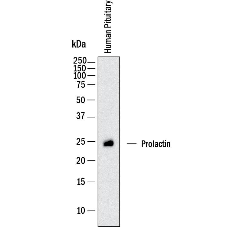
Detection of Human Prolactin by Western Blot. Western blot shows lysates of human pituitary tissue. PVDF membrane was probed with 0.25 µg/mL of Goat Anti-Human Prolactin Antigen Affinity-purified Polyclonal Antibody (Catalog # AF682) followed by HRP-conjugated Anti-Goat IgG Secondary Antibody (Catalog # HAF017). A specific band was detected for Prolactin at approximately 23 kDa (as indicated). This experiment was conducted under reducing conditions and using Immunoblot Buffer Group 1.
 View Larger
Prolactin in Human Testis. Prolactin was detected in immersion fixed paraffin-embedded sections of human testis using Goat Anti-Human Prolactin Antigen Affinity-purified Polyclonal Antibody (Catalog # AF682) at 1 µg/mL overnight at 4 °C. Tissue was stained using the Anti-Goat HRP-DAB Cell & Tissue Staining Kit (brown; Catalog # CTS008) and counterstained with hematoxylin (blue). Specific staining was localized to cytoplasm of sperm cells. View our protocol for Chromogenic IHC Staining of Paraffin-embedded Tissue Sections.
 View Larger
Detection of Human Prolactin by Simple WesternTM. Simple Western shows lysates of human pituitary, loaded at 0.2 mg/ml. A specific band was detected for Prolactin at approximately 30 kDa (as indicated) using 2.5 µg/mL of Goat Anti-Human Prolactin Antigen Affinity-purified Polyclonal Antibody (Catalog # AF682). This experiment was conducted under reducing conditions and using the 12-230kDa separation system.
 View Larger
Neutralization by Human Prolactin Antibody Human Prolactin Antibody (Catalog # AF682) neutralizes Recombinant Human Prolactin (682-PL) induced proliferation in the Nb2 11 rat lymphoma cell line. The Neutralization Dose (ND50) for this effect is typically 0.0200-0.200 µg/mL.
Preparation and Storage
- 12 months from date of receipt, -20 to -70 °C as supplied.
- 1 month, 2 to 8 °C under sterile conditions after reconstitution.
- 6 months, -20 to -70 °C under sterile conditions after reconstitution.
Background: Prolactin
Prolactin (PRL) is a neuroendocrine pituitary hormone. Prolactin is synthesized by the anterior pituitary, placenta, brain, uterus, dermal fibroblasts, decidua, B cell, T cells, NK cells, and breast cancer cells. Originally characterized as a lactogenic hormone, studies have demonstrated broader roles in breast cancer development, regulation of reproductive function, and immunoregulation. In the immune system, prolactin has been shown to be secreted by human PBMC and to act as a proliferative growth factor. Additionally, prolactin treatment of human PBMC has been shown to enhance IFN-gamma production. Prolactin has several molecular forms. The predominant form is a monomer, the non-glycosylated form is 23 kDa and the glycosylated form is 25 kDa. Glycosylated prolactin is removed from the circulation faster and has been reported to have lower biological potency. Prolactin cDNA encodes a 227 amino acid residue protein with a putative 28 aa residue signal peptide. The prolactin receptor is a transmembrane type I glycoprotein that belongs to the cytokine hematopoietic receptor family. B cells, T cells, macrophages, NK cells, monocytes, CD34+ progenitor cells, neutrophils, mammary gland, liver, kidney, adrenals, ovaries, testis, prostrate, seminal vesicles, and hypothalamus have all been shown to express the prolactin receptor. Three forms of the receptor, generated by differential splicing, have been identified. These isoforms differ in the length of their cytoplasmic domains. It is believed that the short cytoplasmic form is non-functional. Prolactin signal transduction involves the JAK/STAT families and Src kinase family.
- Cooke, N.E. et al. (1981) J. Biol. Chem. 256:4007.
- Ben-Johnson, N. et al. (1996) Endoc. Rev. 17:639.
- Cesario, T. et al. (1994) Proc. Soc. Exp. Biol. Med. 205:89.
- Price, A.E. et al. (1995) Endoc. 136:4827.
- Hoffmann, T. et al. (1993) J. Endoc. Invest. 16:807.
- Bellone, G. et al. (1995) J. Cell Physiol. 163:221.
- Cole, E. et al. (1991) Endoc. 129:2639.
- Lewis, U. et al. (1985) Endoc. 116:359.
Product Datasheets
Citations for Human Prolactin Antibody
R&D Systems personnel manually curate a database that contains references using R&D Systems products. The data collected includes not only links to publications in PubMed, but also provides information about sample types, species, and experimental conditions.
7
Citations: Showing 1 - 7
Filter your results:
Filter by:
-
CD36-mediated arachidonic acid influx from decidual stromal cells increases inflammatory macrophages in miscarriage
Authors: Chen, J;Yin, T;Hu, X;Chang, L;Sang, Y;Xu, L;Zhao, W;Liu, L;Xu, C;Lin, Y;Li, Y;Wu, Q;Li, D;Li, Y;Du, M;
Cell reports
Species: Human
Sample Types: Whole Cells
Applications: Neutralization -
The role of Interleukin-18 in recurrent early pregnancy loss
Authors: S Löb, B Ochmann, Z Ma, T Vilsmaier, C Kuhn, E Schmoeckel, SL Herbert, T Kolben, A Wöckel, S Mahner, U Jeschke
Journal of reproductive immunology, 2021-10-05;148(0):103432.
Species: Human
Sample Types: Whole Tissue
Applications: IHC -
Interleukin-1 beta is significantly upregulated in the decidua of spontaneous and recurrent miscarriage placentas
Authors: S Löb, N Amann, C Kuhn, E Schmoeckel, A Wöckel, A Zati Zehni, T Kaltofen, S Keckstein, JN Mumm, S Meister, T Kolben, S Mahner, U Jeschke, T Vilsmaier
Journal of reproductive immunology, 2021-01-30;144(0):103283.
Species: Human
Sample Types: Whole Tissue
Applications: IHC -
Short prolactin isoforms are expressed in photoreceptors of canine retinas undergoing retinal degeneration
Authors: R Sudharsan, L Murgiano, HY Tang, TW Olsen, VRM Chavali, GD Aguirre, WA Beltran
Scientific Reports, 2021-01-11;11(1):460.
Species: Human
Sample Types: Tissue Homogenates
Applications: Western Blot -
Prolactin enhances interferon-gamma-induced production of CXC ligand 9 (CXCL9), CXCL10, and CXCL11 in human keratinocytes.
Authors: Kanda N, Watanabe S
Endocrinology, 2007-01-25;148(5):2317-25.
Species: Human
Sample Types: Cell Culture Supernates
Applications: ELISA Development -
Shift of monocyte function toward cellular immunity during sleep.
Authors: Lange T, Dimitrov S, Fehm HL, Westermann J, Born J
Arch. Intern. Med., 2006-09-18;166(16):1695-700.
Species: Human
Sample Types: Whole Cells
Applications: Neutralization -
Prolactin and heregulin override DNA damage-induced growth arrest and promote phosphatidylinositol-3 kinase-dependent proliferation in breast cancer cells.
Authors: Chakravarti P, Henry MK, Quelle FW
Int. J. Oncol., 2005-02-01;26(2):509-14.
Species: Human
Sample Types: Whole Cells
Applications: Neutralization
FAQs
No product specific FAQs exist for this product, however you may
View all Antibody FAQsReviews for Human Prolactin Antibody
There are currently no reviews for this product. Be the first to review Human Prolactin Antibody and earn rewards!
Have you used Human Prolactin Antibody?
Submit a review and receive an Amazon gift card.
$25/€18/£15/$25CAN/¥75 Yuan/¥2500 Yen for a review with an image
$10/€7/£6/$10 CAD/¥70 Yuan/¥1110 Yen for a review without an image
