Human TRIM21 Antibody Summary
Met1-Tyr475
Accession # P19474
Applications
Please Note: Optimal dilutions should be determined by each laboratory for each application. General Protocols are available in the Technical Information section on our website.
Scientific Data
 View Larger
Detection of Human TRIM21 by Western Blot. Western blot shows lysates of HeLa human cervical epithelial carcinoma cell line and human kidney tissue. PVDF Membrane was probed with 0.5 µg/mL of Human TRIM21 Antigen Affinity-purified Polyclonal Antibody (Catalog # AF6219) followed by HRP-conjugated Anti-Sheep IgG Secondary Antibody (HAF016). A specific band was detected for TRIM21 at approximately 52 kDa (as indicated). This experiment was conducted under reducing conditions and using Immunoblot Buffer Group 2.
 View Larger
TRIM21 in Human PBMCs. TRIM21 was detected in immersion fixed human peripheral blood mononuclear cells (PBMCs) stimulated for 8 hours with 20 ng/mL Recombinant Human IFN-gamma (Catalog # 285-IF) using Human TRIM21 Antigen Affinity-purified Polyclonal Antibody (Catalog # AF6219) at 10 µg/mL for 3 hours at room temperature. Cells were stained using the NorthernLights™ 557-conjugated Anti-Sheep IgG Secondary Antibody (red; NL010) and counterstained with DAPI (blue). Specific staining was localized to cytoplasm. View our protocol for Fluorescent ICC Staining of Non-adherent Cells.
 View Larger
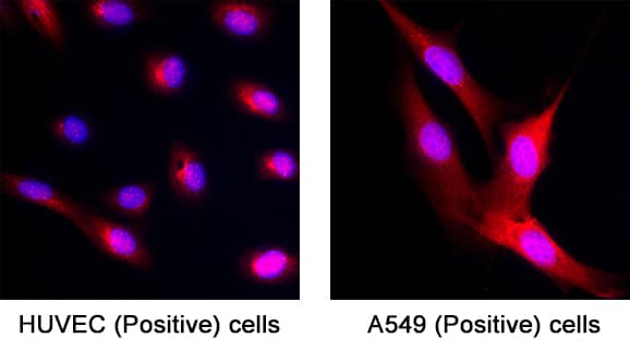
Detection of TRIM21 in HUVEC Human Umbilical Vein Endothelial Cells and A549 Human Lung Carcinoma Cells (Both Positive Controls). TRIM21 was detected in immersion fixed HUVEC Human Umbilical Vein Endothelial Cells and A549 Human Lung Carcinoma Cells (Both Positive Controls) using Sheep Anti-Human TRIM21 Antigen Affinity-purified Polyclonal Antibody (Catalog # AF6219) at 15 µg/mL for 3 hours at room temperature. Cells were stained using the NorthernLights™ 557-conjugated Anti-Sheep IgG Secondary Antibody (red; Catalog # NL010) and counterstained with DAPI (blue). Specific staining was localized to cytoplasm and cell nuclei. View our protocol for Fluorescent ICC Staining of Cells on Coverslips.
Preparation and Storage
- 12 months from date of receipt, -20 to -70 °C as supplied.
- 1 month, 2 to 8 °C under sterile conditions after reconstitution.
- 6 months, -20 to -70 °C under sterile conditions after reconstitution.
Background: TRIM21
TRIM21 (Tripartite motif-containing protein 21; also Ro(SS-A), 52 kDa Ro Protein/Ro52, and RING finger protein 81) is a 52-56 kDa member of the RING finger B box coiled coil family of proteins. It is an E3 ligase that is found in both nucleus and cytoplasm, where it is often associated with microtubules. TRIM21 ubiquitinates select proteins. In B cells, it targets the Fc fragment of misfolded IgG, providing QC on its production. In macrophages, it acts in a nondegradative manner on IRF8, promoting innate immunity. Human TRIM21 is 475 amino acids (aa) in length and contains one E3 ligase RING finger domain (aa 16-55), a B Box type zinc finger region (aa 92-123), a coiled coil region (aa 128-238) and a C-terminal SPRY/B30.2 Ig binding domain (aa 268-465). TRIM21 is reported to form trimers. Over aa 195‑293, human TRIM21 exhibits 72% aa identity with mouse TRIM21.
Product Datasheets
FAQs
No product specific FAQs exist for this product, however you may
View all Antibody FAQsIsotype Controls
Reconstitution Buffers
Secondary Antibodies
Reviews for Human TRIM21 Antibody
There are currently no reviews for this product. Be the first to review Human TRIM21 Antibody and earn rewards!
Have you used Human TRIM21 Antibody?
Submit a review and receive an Amazon gift card.
$25/€18/£15/$25CAN/¥75 Yuan/¥2500 Yen for a review with an image
$10/€7/£6/$10 CAD/¥70 Yuan/¥1110 Yen for a review without an image
