Mouse Mer APC-conjugated Antibody Summary
Glu23-Phe498
Accession # Q60805
Applications
Please Note: Optimal dilutions should be determined by each laboratory for each application. General Protocols are available in the Technical Information section on our website.
Scientific Data
 View Larger
Detection of Mer in J774A.1 Mouse Cell Line by Flow Cytometry. J774A.1 mouse reticulum cell sarcoma macrophage cell line was stained with Rat Anti-Mouse Mer APC-conjugated Monoclonal Antibody (Catalog # FAB5912A, filled histogram) or isotype control antibody (Catalog # IC006A, open histogram). View our protocol for Staining Membrane-associated Proteins.
Reconstitution Calculator
Preparation and Storage
- 12 months from date of receipt, 2 to 8 °C as supplied.
Background: Mer
Axl (Ufo, Ark), Dtk (Sky, Tyro3, Rse, Brt) and Mer (human and mouse homologues of chicken c-Eyk) constitute a receptor tyrosine kinase subfamily. The extracellular domains of these proteins contain two Ig-like motifs and two fibronectin type III motifs. This characteristic topology is also found in neural cell adhesion molecules and in receptor tyrosine phosphatases. These receptors bind the vitamin K-dependent protein Growth-Arrest-Specific gene 6 (Gas6) which is structurally related to the anticoagulation factor protein S. Binding of Gas6 induces receptor autophosphorylation and downstream signaling pathways that can lead to cell proliferation, migration or the prevention of apoptosis. Studies suggest that this family of tyrosine kinase receptors may be involved in hematopoiesis, embryonic development, tumorigenesis and regulation of testicular functions (1-2).
- Nagata, K. et al. (1996) J. Biol. Chem. 22:30022.
- Crosier, K.E. and P.S Crosier (1997) Pathology 29:131.
Product Datasheets
Citations for Mouse Mer APC-conjugated Antibody
R&D Systems personnel manually curate a database that contains references using R&D Systems products. The data collected includes not only links to publications in PubMed, but also provides information about sample types, species, and experimental conditions.
7
Citations: Showing 1 - 7
Filter your results:
Filter by:
-
Isolevuglandins disrupt PU.1-mediated C1q expression and promote autoimmunity and hypertension in systemic lupus erythematosus
Authors: DM Patrick, N de la Visi, J Krishnan, W Chen, MJ Ormseth, CM Stein, SS Davies, V Amarnath, LJ Crofford, JM Williams, S Zhao, CD Smart, S Dikalov, A Dikalova, L Xiao, JP Van Beusec, M Ao, AB Fogo, A Kirabo, DG Harrison
JCI Insight, 2022-07-08;7(13):.
Species: Mouse
Sample Types: Whole Cells
Applications: Flow Cytometry -
Cardiac Mesenchymal Stem Cells Promote Fibrosis and Remodeling in Heart Failure: Role of PDGF Signaling
Authors: Tariq Hamid, Yuanyuan Xu, Mohamed Ameen Ismahil, Gregg Rokosh, Miki Jinno, Guihua Zhou et al.
JACC: Basic to Translational Science
-
IFN-beta is a macrophage-derived effector cytokine facilitating the resolution of bacterial inflammation
Authors: Senthil Kumaran Satyanarayanan, Driss El Kebir, Soaad Soboh, Sergei Butenko, Meriem Sekheri, Janan Saadi et al.
Nature Communications
-
Dectin-2-induced CCL2 production in tissue-resident macrophages ignites cardiac arteritis
Authors: C Miyabe, Y Miyabe, L Moreno, J Lian, RA Rahimi, NN Miura, N Ohno, Y Iwakura, T Kawakami, AD Luster
J. Clin. Invest., 2019-06-06;130(0):.
Species: Mouse
Sample Types: Whole Cells
Applications: Flow Cytometry -
Csf1r or Mer inhibition delays liver regeneration via suppression of Kupffer cells
Authors: JA Santamaria, S Zeng, JB Greer, MJ Beckman, AM Seifert, NA Cohen, JQ Zhang, MH Crawley, BL Green, JK Loo, JH Maltbaek, RP DeMatteo
PLoS ONE, 2019-05-01;14(5):e0216275.
Species: Mouse
Sample Types: Whole Cells
Applications: Flow Cytometry -
MerTK cleavage limits proresolving mediator biosynthesis and exacerbates tissue inflammation
Proc Natl Acad Sci USA, 2016-05-19;0(0):.
Species: Mouse
Sample Types: Whole Cells
Applications: Flow Cytometry -
The monocyte to macrophage transition in the murine sterile wound.
Authors: Crane, Meredith, Daley, Jean M, van Houtte, Olivier, Brancato, Samielle, Henry, William, Albina, Jorge E
PLoS ONE, 2014-01-22;9(1):e86660.
Species: Mouse
Sample Types: Whole Cells
Applications: Flow Cytometry
FAQs
No product specific FAQs exist for this product, however you may
View all Antibody FAQsReviews for Mouse Mer APC-conjugated Antibody
Average Rating: 4.7 (Based on 3 Reviews)
Have you used Mouse Mer APC-conjugated Antibody?
Submit a review and receive an Amazon gift card.
$25/€18/£15/$25CAN/¥75 Yuan/¥2500 Yen for a review with an image
$10/€7/£6/$10 CAD/¥70 Yuan/¥1110 Yen for a review without an image
Filter by:
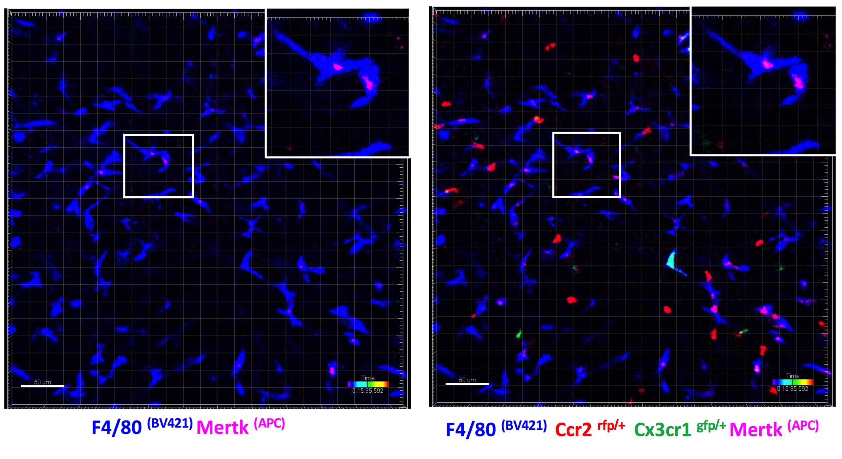
In vivo imaging of liver macrophages using intravital laser confocal microscopy. Ccr2/Cx3cr1 transgenic mice were anaesthetised with ketamine and medetomidine, received i.v. injection of fluorophore-conjugated antibodies [anti-F4/80 (BV421) and anti-MerTK (APC)] and then surgical exposure of the liver was performed. Imaging was performed under isofluorane anaesthesia on a Leica SP5 laser scanning confocal microscope using a 20x objective. Videos were recorded at defined regions of interest. Image analysis was performed using Imaris (v 8.0). Representative screen shots of intravital liver imaging videos showing F4/80+ macrophages (blue) expressing MerTK (pink), ccr2 (red) or cx3cr1 (green), located in centrilobular areas of the liver.

