Mouse NCAM-1/CD56 Antibody Summary
Leu20-Thr711
Accession # P13595
Applications
Please Note: Optimal dilutions should be determined by each laboratory for each application. General Protocols are available in the Technical Information section on our website.
Scientific Data
 View Larger
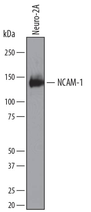
Detection of Mouse NCAM‑1/CD56 by Western Blot. Western blot shows lysates of Neuro-2A mouse neuroblastoma cell line. PVDF membrane was probed with 0.1 µg/mL of Rat Anti-Mouse NCAM-1/CD56 Monoclonal Antibody (Catalog # MAB7820) followed by HRP-conjugated Anti-Rat IgG Secondary Antibody (Catalog # HAF005). A specific band was detected for NCAM-1/CD56 at approximately 120 kDa (as indicated). This experiment was conducted under reducing conditions and using Immunoblot Buffer Group 1.
 View Larger
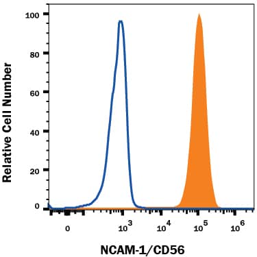
Detection of NCAM‑1/CD56 in Neuro‑2A Mouse Cell Line by Flow Cytometry. Neuro-2A mouse neuroblastoma cell line was stained with Rat Anti-Mouse NCAM-1/CD56 Monoclonal Antibody (Catalog # MAB7820, filled histogram) or isotype control antibody (Catalog # MAB006, open histogram), followed by Allophycocyanin-conjugated Anti-Rat IgG Secondary Antibody (Catalog # F0113). View our protocol for Staining Membrane-associated Proteins.
 View Larger
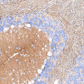
Detection of NCAM‑1/CD56 in Mouse Cerebellum. NCAM‑1/CD56 was detected in immersion fixed paraffin-embedded sections of mouse cerebellum using Rat Anti-Mouse NCAM‑1/CD56 Monoclonal Antibody (Catalog # MAB7820) at 5 µg/ml for 1 hour at room temperature followed by incubation with the Anti-Rat IgG VisUCyte™ HRP Polymer Antibody (Catalog # VC005). Before incubation with the primary antibody, tissue was subjected to heat-induced epitope retrieval using VisUCyte Antigen Retrieval Reagent-Basic (Catalog # VCTS021). Tissue was stained using DAB (brown) and counterstained with hematoxylin (blue). Specific staining was localized to the cytoplasm and neuropils. View our protocol for Chromogenic IHC Staining of Paraffin-embedded Tissue Sections.
 View Larger
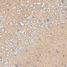
Detection of NCAM‑1/CD56 in Mouse Cortex. NCAM‑1/CD56 was detected in immersion fixed paraffin-embedded sections of mouse cortex using Rat Anti-Mouse NCAM‑1/CD56 Monoclonal Antibody (Catalog # MAB7820) at 5 µg/ml for 1 hour at room temperature followed by incubation with the Anti-Rat IgG VisUCyte™ HRP Polymer Antibody (Catalog # VC005). Before incubation with the primary antibody, tissue was subjected to heat-induced epitope retrieval using VisUCyte Antigen Retrieval Reagent-Basic (Catalog # VCTS021). Tissue was stained using DAB (brown) and counterstained with hematoxylin (blue). Specific staining was localized to the cytoplasm and neuropils. View our protocol for Chromogenic IHC Staining of Paraffin-embedded Tissue Sections.
Preparation and Storage
- 12 months from date of receipt, -20 to -70 °C as supplied.
- 1 month, 2 to 8 °C under sterile conditions after reconstitution.
- 6 months, -20 to -70 °C under sterile conditions after reconstitution.
Background: NCAM-1/CD56
NCAM-1 (Neural adhesion molecule-1; also CD56) is a 120-190 kDa glycoprotein member of the Ig Superfamily. It is expressed on multiple cell types, both in the embryo and adult. Here, it serves as both an adhesion molecule and a receptor for multiple ligands, including as FGFR, PDGF, GDNF and agrin. On the cell surface, it is a cis-oriented homodimer that can form homodimers in-trans with other cis-homodimers. In the embryo, NCAM-1 is polysialylated (PolySia), and shows a MW of 200-220 kDa in SDS-PAGE. This polysialylation reduces the ability of NCAM-1 to dimerize. Mature mouse NCAM-1 is a 1096 amino acid (aa) type I transmembrane (TM) protein (aa 20-1115). It possesses a 692 aa extracellular region (aa 20-711) and a 386 aa cytoplasmic domain. The extracellular region contains five consecutive C2-type Ig-like domains (aa 20-492) followed by two FN type-III domains (aa 497-692). Multiple splice variants exist. There is a 140 kDa TM variant that shows a deletion of aa 810-1076, and a 120 kDa variant that is GPI-linked and shows a 24 aa substitution for aa 702-1115. A third potential variant contains a five aa substitution for aa 601-1115. Over aa 20-711, mouse NCAM-1 shares 99% and 95% aa identity with rat and human NCAM-1, respectively.
Product Datasheets
Citations for Mouse NCAM-1/CD56 Antibody
R&D Systems personnel manually curate a database that contains references using R&D Systems products. The data collected includes not only links to publications in PubMed, but also provides information about sample types, species, and experimental conditions.
2
Citations: Showing 1 - 2
Filter your results:
Filter by:
-
Diminished expression of major histocompatibility complex facilitates the use of human induced pluripotent stem cells in monkey
Authors: X Wang, M Lu, X Tian, Y Ren, Y Li, M Xiang, S Chen
Stem Cell Res Ther, 2020-08-03;11(1):334.
Species: Human
Sample Types: Cell Lysates
Applications: Western Blot -
The prostaglandin E2 receptor EP3 controls CC-chemokine ligand-mediated neuropathic pain induced by mechanical nerve damage
Authors: EM Treutlein, K Kern, A Weigert, N Tarighi, CD Schuh, RM Nüsing, Y Schreiber, N Ferreirós, B Brüne, G Geisslinge, S Pierre, K Scholich
J. Biol. Chem., 2018-05-11;0(0):.
Species: Mouse
Sample Types: Whole Cells
Applications: Flow Cytometry
FAQs
No product specific FAQs exist for this product, however you may
View all Antibody FAQsReviews for Mouse NCAM-1/CD56 Antibody
There are currently no reviews for this product. Be the first to review Mouse NCAM-1/CD56 Antibody and earn rewards!
Have you used Mouse NCAM-1/CD56 Antibody?
Submit a review and receive an Amazon gift card.
$25/€18/£15/$25CAN/¥75 Yuan/¥2500 Yen for a review with an image
$10/€7/£6/$10 CAD/¥70 Yuan/¥1110 Yen for a review without an image


