Phospho-Akt (S473) PE-conjugated Antibody Summary
Applications
Please Note: Optimal dilutions should be determined by each laboratory for each application. General Protocols are available in the Technical Information section on our website.
Scientific Data
 View Larger
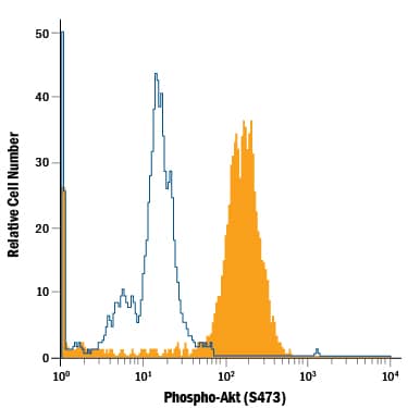
Detection of Phospho-Akt (S473) in Jurkat Human Cell Line by Flow Cytometry. Jurkat human acute T cell leukemia cell line either untreated (open histogram) or treated with 100 nM Calyculin A (Catalog # 1336) for 30 minutes (filled histogram) was stained with Mouse Anti-Human/Mouse Phospho-Akt (S473) PE-conjugated Monoclonal Antibody (Catalog # IC7794P). To facilitate intracellular staining, cells were fixed with Flow Cytometry Fixation Buffer (Catalog # FC004) and permeabilized with Flow Cytometry Permeabilization/Wash Buffer I (Catalog # FC005). View our protocol for Staining Intracellular Molecules.
Reconstitution Calculator
Preparation and Storage
- 12 months from date of receipt, 2 to 8 °C as supplied.
Background: Akt
Akt, also known as Protein Kinase B (PKB), is a central kinase in such diverse cellular processes as glucose uptake, cell cycle progression, and apoptosis. Three highly homologous members define the Akt family: Akt1 (PKB alpha ), Akt2 (PKB beta ), and Akt3 (PKB gamma ). All three Akts contain an amino-terminal pleckstrin homology domain, a central kinase domain, and a carboxyl-terminal regulatory domain. Akt1 is the most widely expressed family member and is frequently activated in a number of carcinomas, including breast, prostate, lung, pancreatic, liver, ovarian, and colorectal cancer. Akt1 is activated in a multistep process that involves the sequential phosphorylation of Thr450 by JNK kinases, Thr308 by PDK1, and Ser473 by PDK2 or mTORC2. Activated Akt1 phosphorylates a wide variety of cytosolic, nuclear, and mitochondrial substrates. Human Akt1 shares 98% aa sequence identity with mouse and rat Akt1.
Product Datasheets
FAQs
No product specific FAQs exist for this product, however you may
View all Antibody FAQsReviews for Phospho-Akt (S473) PE-conjugated Antibody
Average Rating: 4 (Based on 1 Review)
Have you used Phospho-Akt (S473) PE-conjugated Antibody?
Submit a review and receive an Amazon gift card.
$25/€18/£15/$25CAN/¥75 Yuan/¥2500 Yen for a review with an image
$10/€7/£6/$10 CAD/¥70 Yuan/¥1110 Yen for a review without an image
Filter by:


