Organelles: Endosomes
Intracellular Junctions
Nuclear Envelope
Nucleus
Endoplasmic Reticulum
Ribosomes
Golgi Apparatus
Endosomes
Mitochondria
Plasma Membrane
Lysosomes
Autophagosomes
Peroxisomes
Cytoskeleton
View Organelles Main

Caveolin-1
Caveolin-1
ProductsClose
Caveolin-2
Caveolin-2
ProductsClose
Clathrin
Heavy
Chain 1/
CHC17
Heavy
Chain 1/
CHC17
Clathrin
Heavy
Chain 1/
CHC17
Heavy
Chain 1/
CHC17
ProductsClose
Clathrin
Heavy
Chain 2/
CHC22
Heavy
Chain 2/
CHC22
Clathrin
Heavy
Chain 2/
CHC22
Heavy
Chain 2/
CHC22
ProductsClose
DAB2
DAB2
EEA1
EEA1
IGF-II R
IGF-II R
PAK1
PAK1
Rab27a
Rab27a
Rab7a
Rab7a
Syntaxin 7
Syntaxin 7
ProductsClose
TfR (Transferrin R)
TfR (Transferrin R)
ProductsClose
Products
Products
Overview
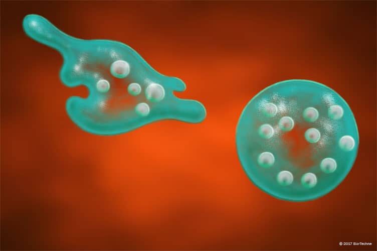
Endosomes are derived from the plasma membrane during endocytosis. They mediate cellular uptake and plasma membrane recycling by fusing with the Golgi secretory pathway. They can also target select membrane components for lysosomal degradation.
Data Examples
Caveolin-1 in HeLa Human Cell Line.
Caveolin-1 was detected in formaldehyde fixed HeLa human cervical epithelial carcinoma cell line using Mouse Anti-Human/Mouse/Rat Caveolin-1 Biotinylated Monoclonal Antibody (Catalog # BAM5736) at 25 µg/mL overnight at 4 °C. Cells were stained using the NorthernLights™ 493-conjugated Streptavidin (green; Catalog # NL997) and counterstained with DAPI (blue). Specific staining was localized to caveolae. View our protocol for Fluorescent ICC Staining of Cells on Coverslips.
EEA1 in HeLa Human Cell Line. EEA1 was detected in formaldehyde fixed HeLa human cervical epithelial carcinoma cell line using Sheep Anti-Human/Mouse/Rat EEA1 Biotinylated Antigen Affinity-purified Polyclonal Antibody (Catalog # BAF8047) at 15 µg/mL overnight at 4°C. Cells were stained using the NorthernLights™ 557-conjugated Streptavidin (orange; Catalog # NL999) and counterstained with DAPI (blue). Specific staining was localized to endosomes. View our protocol for Fluorescent ICC Staining of Cells on Coverslips.
Detection of Human and Mouse Rab7a by Western Blot. Western blot shows lysates of SK-Mel-28 human malignant melanoma cell line and B16-F1 mouse melanoma cell line. PVDF membrane was probed with 1 µg/mL of Sheep Anti-Human/Mouse Rab7a Antigen Affinity-purified Polyclonal Antibody (Catalog # HAF016). A specific band was detected for Rab7a at approximately 23 kDa (as indicated). This experiment was conducted under reducing conditions and using Immunoblot Buffer Group 1.


