Human/Mouse Semaphorin 3C Antibody Summary
Gln24-Ser751 (Arg548Ala, Arg552Ala)
Accession # Q62181
*Small pack size (-SP) is supplied either lyophilized or as a 0.2 µm filtered solution in PBS.
Customers also Viewed
Applications
Please Note: Optimal dilutions should be determined by each laboratory for each application. General Protocols are available in the Technical Information section on our website.
Scientific Data
 View Larger
Semaphorin 3C in MCF‑7 Human Cell Line. Semaphorin 3C was detected in immersion fixed MCF-7 human breast cancer cell line using Rat Anti-Mouse Semaphorin 3C Monoclonal Antibody (Catalog # MAB1728) at 10 µg/mL for 3 hours at room temperature. Cells were stained using the NorthernLights™ 557-conjugated Anti-Rat IgG Secondary Antibody (red; Catalog # NL013) and counterstained with DAPI (blue). Specific staining was localized to cytoplasm. View our protocol for Fluorescent ICC Staining of Cells on Coverslips.
 View Larger
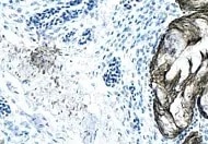
Semaphorin 3C in Mouse Embryo. Semaphorin 3C was detected in immersion fixed frozen sections of mouse embryo (13 d.p.c.) using Rat Anti-Human/Mouse Semaphorin 3C Monoclonal Antibody (Catalog # MAB1728) at 1.7 µg/mL overnight at 4 °C. Tissue was stained using the Anti-Rat HRP-DAB Cell & Tissue Staining Kit (brown; Catalog # CTS017) and counterstained with hematoxylin (blue). Specific staining was localized to developing muscle cells. View our protocol for Chromogenic IHC Staining of Frozen Tissue Sections.
Preparation and Storage
- 12 months from date of receipt, -20 to -70 °C as supplied.
- 1 month, 2 to 8 °C under sterile conditions after reconstitution.
- 6 months, -20 to -70 °C under sterile conditions after reconstitution.
Background: Semaphorin 3C
Semaphorin 3C (Sema3C; previously semaE) is one of six Class 3 secreted semaphorins which share 40-50% amino acid (aa) identity. Class 3 semaphorins are potent chemorepellents that function in axon and/or vascular guidance during development, and may be upregulated in tumor progression (1, 2). The 751 amino acid (aa) mouse Sema3C is highly modular. It contains a 20 aa signal sequence, an ~500 aa N-terminal Sema domain that forms a beta -propeller structure similar to that found in integrin molecules, a cysteine knot, a furin-type cleavage site, an Ig-like domain, and a C-terminal basic domain (1-3). Covalent dimerization plus cleavage at the C-terminus are required for activity of class 3 semaphorins (4). Mouse Sema3C shares at least 95% aa identity with human, rat, cow and dog Sema3C, and 89% and 75% aa identity with chick and zebrafish Sema3C, respectively. Type 3 semaphorins transduce signals through transmembrane plexins, either directly or by binding associated neuropilin receptors (1, 2). Sema3C signaling is transduced by Plexin-D1 indirectly via neuropilin-1 or neuropilin-2 receptors (5). Sema3C is expressed in all somitic motor neurons, in lung buds and in cardiac neural crest cells during development (1, 5-8). Sema3C activates integrins in certain cells so, in addition to its repulsive activities, it sometimes acts as a chemoattractant (6, 9). In the developing nervous system, this chemoattraction appears to complement Sema3A repulsion in adjacent cell layers (1, 6, 7). Sema3C also provides an attractive force opposing Sema6A and Sema6B to guide migration of neural crest endothelial cells to the cardiac outflow tract (10). Consequently, defects in aortic arch formation occur when Sema3C or Plexin-D1 genes or Sema3C-neuropilin interactions are disrupted (5, 11, 12).
- Hinck, L. (2004) Dev. Cell 7:783.
- Neufeld, G. et al. (2005) Front. Biosci. 10:751.
- Gherardi, E. et al. (2004) Curr. Opin. Struct. Biol. 14:669.
- Adams, R. H. et al. (1997) EMBO J. 16:6077.
- Gitler, A. D. et al. (2004) Dev. Cell 7:107.
- Bagnard, D. et al. (1998) Development 125:5043.
- Cohen, S. et al. (2005) Eur. J. Neurosci. 21:1767.
- Puschel, A. W. et al. (1995) Neuron 14:941.
- Herman, J. G. and G. G. Meadows (2007) Int. J. Oncol. 30:1231.
- Toyofuku, T. et al. (2008) Dev. Biol. 321:251.
- Feiner, L. et al. (2001) Development 128:3061.
- Gu, C. et al. (2003) Dev. Cell 5:45.
Product Datasheets
Citations for Human/Mouse Semaphorin 3C Antibody
R&D Systems personnel manually curate a database that contains references using R&D Systems products. The data collected includes not only links to publications in PubMed, but also provides information about sample types, species, and experimental conditions.
7
Citations: Showing 1 - 7
Filter your results:
Filter by:
-
Integrated single-cell analyses decode the developmental landscape of the human fetal spine
Authors: Yu H, Tang D, Wu H et al.
iScience
-
Sema3C signaling is an alternative activator of the canonical WNT pathway in glioblastoma
Authors: J Hao, X Han, H Huang, X Yu, J Fang, J Zhao, RA Prayson, S Bao, JS Yu
Nature Communications, 2023-04-20;14(1):2262.
Species: Human
Sample Types: Cell Lysates
Applications: Western Blot -
Motor neurons use push-pull signals to direct vascular remodeling critical for their connectivity
Authors: Luis F. Martins, Ilaria Brambilla, Alessia Motta, Stefano de Pretis, Ganesh Parameshwar Bhat, Aurora Badaloni et al.
Neuron
Species: Monkey
Sample Types: Cell Lysates
Applications: Western Blot -
The cleavage of semaphorin 3C induced by ADAMTS1 promotes cell migration.
Authors: Esselens C, Malapeira J, Colome N, Casal C, Rodriguez-Manzaneque JC, Canals F, Arribas J
J. Biol. Chem., 2009-11-13;285(4):2463-73.
Species: Human
Sample Types: Cell Lysates
Applications: Western Blot -
Motor neurons use push-pull signals to direct vascular remodeling critical for their connectivity
Authors: Luis F. Martins, Ilaria Brambilla, Alessia Motta, Stefano de Pretis, Ganesh Parameshwar Bhat, Aurora Badaloni et al.
Neuron
-
Developing Potential Candidates of Preclinical Preeclampsia
Authors: Sandra Founds, Xuemei Zeng, David Lykins, James M. Roberts
International Journal of Molecular Sciences
-
SEMA3C Promotes Cervical Cancer Growth and Is Associated With Poor Prognosis
Authors: Ruoyan Liu, Yanjie Shuai, Jingtao Luo, Ze Zhang
Frontiers in Oncology
FAQs
No product specific FAQs exist for this product, however you may
View all Antibody FAQsIsotype Controls
Reconstitution Buffers
Secondary Antibodies
Reviews for Human/Mouse Semaphorin 3C Antibody
Average Rating: 4.5 (Based on 2 Reviews)
Have you used Human/Mouse Semaphorin 3C Antibody?
Submit a review and receive an Amazon gift card.
$25/€18/£15/$25CAN/¥75 Yuan/¥2500 Yen for a review with an image
$10/€7/£6/$10 CAD/¥70 Yuan/¥1110 Yen for a review without an image
Filter by:
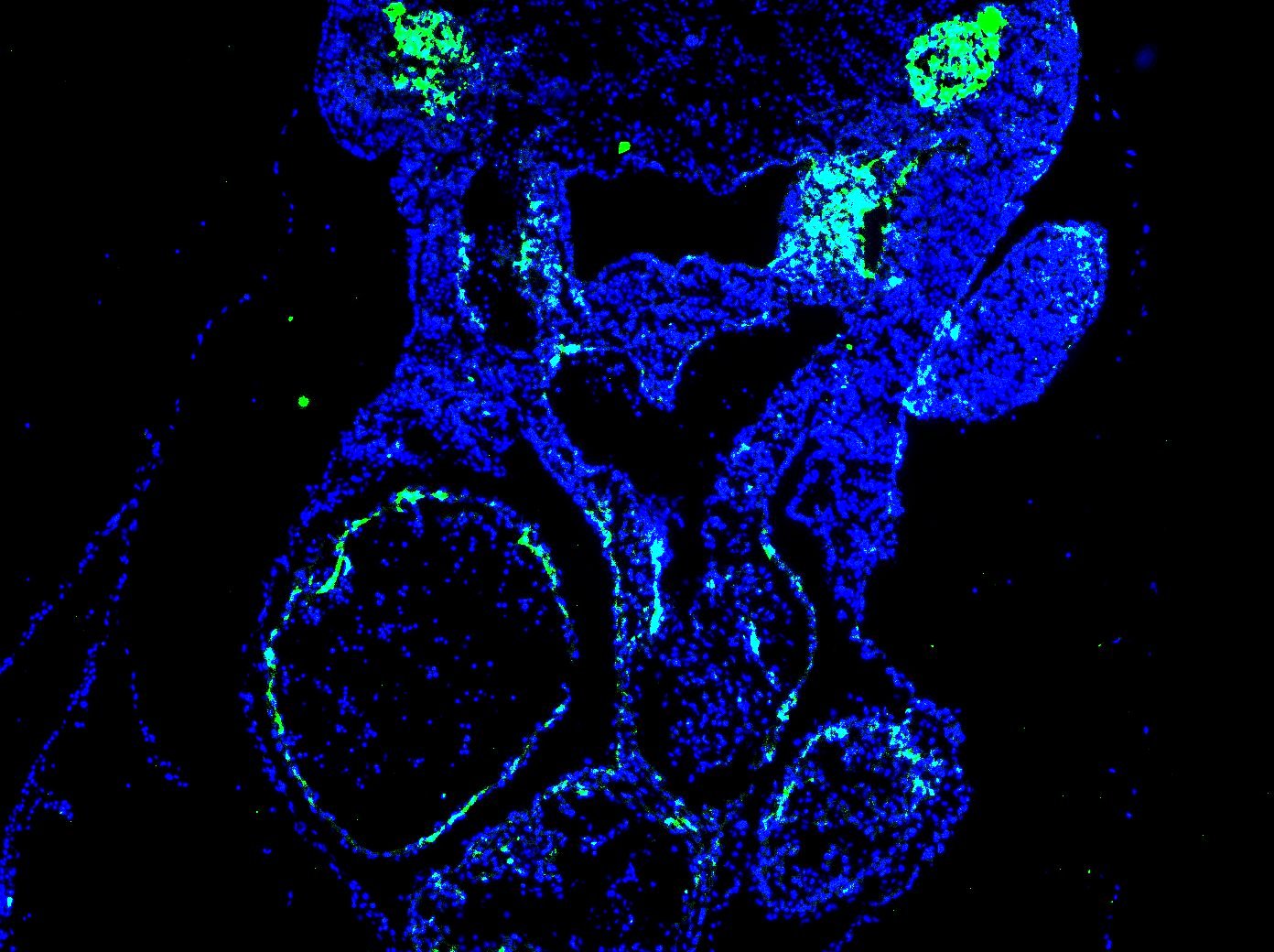
Dilution used - 1:200. The staining was done on an E10.5 mouse transverse section (fixed on 4% PFA overnight) and done using standard IF techniques.
The staining was very good on the target region (look at the upper region of picture which shows excellent Semaphorin3C staining on supposed neural crest cells).
However, there's still a little non specific staining on the arterial lining of the heart, which is a small cause of concern.
Probably this could be eliminated by tweaking the fixation/blocking steps.


