Human SHP-1 Antibody Summary
Ala205-Lys595
Accession # P29350
Customers also Viewed
Applications
Please Note: Optimal dilutions should be determined by each laboratory for each application. General Protocols are available in the Technical Information section on our website.
Scientific Data
 View Larger
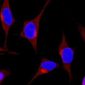
SHP‑1 in HeLa Human Cell Line. SHP-1 was detected in immersion fixed HeLa human cervical epithelial carcinoma cell line using Rat Anti-Human SHP-1 Monoclonal Antibody (Catalog # MAB1878) at 3 µg/mL for 3 hours at room temperature. Cells were stained using the NorthernLights™ 557-conjugated Anti-Rat IgG Secondary Antibody (red; Catalog # NL013) and counterstained with DAPI (blue). Specific staining was localized to cytoplasm. View our protocol for Fluorescent ICC Staining of Cells on Coverslips.
Preparation and Storage
- 12 months from date of receipt, -20 to -70 °C as supplied.
- 1 month, 2 to 8 °C under sterile conditions after reconstitution.
- 6 months, -20 to -70 °C under sterile conditions after reconstitution.
Background: SHP-1
Src-Homology 2 domain Phosphatase-1 (SHP-1), also known as Protein Tyrosine Phosphatase 1C (PTP1C), PTPN6, and Hematopoetic Cell Phosphatase (HCPH), is an enzyme that selectively dephosphorylates tyrosine residues in proteins. Spontaneous point mutations in the SHP-1 gene in mice produce the “motheaten” and "motheaten viable” phenotypes that are severely autoimmune and immunodeficient (1). The enzyme is highly expressed in leukocyte cell types (2). SHP-1 has a regulatory region containing two Src homology 2 (SH2) domains that are critical for its binding to ITIM domains in inhibitory immunoreceptors (3). Deletion of the SH2 domains, as in this product, causes a marked increase in phosphatase activity (4). SHP-1 will dephosphorylate a wide variety of proteins, including the EGF receptor (5). A phosphopeptide containing the EGFR (Y992) sequence (R&D Systems, Catalog # ES006) can be used to measure the activity of SHP-1 by detecting the release of phosphate (R&D Systems, Catalog # DY996).
- Tsui, H.W. et al. (1993) Nature Genet. 4:124.
- Matthews, R.J. et al. (1992) Mol. Cell. Biol. 12:2396.
- Burshtyn, D.N. et al. (1997) J. Biol. Chem. 272:13066.
- Pei, D. et al. (1994) Biochemistry 33:15483.
- Tomic, S. et al. (1995) J. Biol. Chem. 270:21277.
Product Datasheets
Citations for Human SHP-1 Antibody
R&D Systems personnel manually curate a database that contains references using R&D Systems products. The data collected includes not only links to publications in PubMed, but also provides information about sample types, species, and experimental conditions.
2
Citations: Showing 1 - 2
Filter your results:
Filter by:
-
The oncoprotein gankyrin promotes the development of colitis-associated cancer through activation of STAT3
Authors: T Sakurai, H Higashitsu, H Kashida, T Watanabe, Y Komeda, T Nagai, S Hagiwara, M Kitano, N Nishida, T Abe, H Kiyonari, K Itoh, J Fujita, M Kudo
Oncotarget, 2017-04-11;8(15):24762-24776.
Species: Mouse
Sample Types: Whole Tissue
Applications: IHC -
Regulation and function of immunosuppressive molecule human leukocyte antigen G5 in human bone tissue.
Authors: Deschaseaux F, Gaillard J, Langonne A, Chauveau C, Naji A, Bouacida A, Rosset P, Heymann D, De Pinieux G, Rouas-Freiss N, Sensebe L
FASEB J, 2013-04-16;27(8):2977-87.
Species: Human
Sample Types: Whole Cells
Applications: ChIP
FAQs
No product specific FAQs exist for this product, however you may
View all Antibody FAQsIsotype Controls
Reconstitution Buffers
Secondary Antibodies
Reviews for Human SHP-1 Antibody
There are currently no reviews for this product. Be the first to review Human SHP-1 Antibody and earn rewards!
Have you used Human SHP-1 Antibody?
Submit a review and receive an Amazon gift card.
$25/€18/£15/$25CAN/¥75 Yuan/¥2500 Yen for a review with an image
$10/€7/£6/$10 CAD/¥70 Yuan/¥1110 Yen for a review without an image


