Human CD19 Antibody Summary
Met1-Lys291
Accession # P15391
Customers also Viewed
Applications
Please Note: Optimal dilutions should be determined by each laboratory for each application. General Protocols are available in the Technical Information section on our website.
Scientific Data
 View Larger
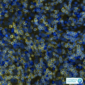
Detection of CD19 in Human Lymph Node. CD19 was detected in immersion fixed paraffin-embedded sections of Human Lymph Node using Mouse Anti-Human CD19 Monoclonal Antibody (Catalog # MAB48671) at 5 µg/mL for 1 hour at room temperature followed by incubation with the Anti-Mouse IgG VisUCyte™ HRP Polymer Antibody (Catalog # VC001). Before incubation with the primary antibody, tissue was subjected to heat-induced epitope retrieval using VisUCyte Antigen Retrieval Reagent-Basic (Catalog # VCTS021). Tissue was stained using DAB (brown) and counterstained with hematoxylin (blue). Specific staining was localized to cell surface in lymphocytes. View our protocol for IHC Staining with VisUCyte HRP Polymer Detection Reagents.
 View Larger
Detection of CD19 in Daudi cells (positive) and RPMI 8226 cells (negative). CD19 was detected in immersion fixed Daudi human Burkitt's lymphoma cell line (positive) and absent in RPMI 8226 human multiple myeloma cell line (negative) using Mouse Anti-Human CD19 Monoclonal Antibody (Catalog # MAB48671) at 8 µg/mL for 3 hours at room temperature. Cells were stained using the NorthernLights™ 557-conjugated Anti-Mouse IgG Secondary Antibody (red; Catalog # NL007) and counterstained with DAPI (blue). Specific staining was localized to cell surface. View our protocol for Fluorescent ICC Staining of Non-adherent Cells.
 View Larger
Detection of CD19 in Daudi cell line (positive) and U266 cell line (negative). CD19 was detected in immersion fixed Daudi human Burkitt's lymphoma cell line (positive) and absent in U266 human myeloma cell line (negative) using Mouse Anti-Human CD19 Monoclonal Antibody (Catalog # MAB48671) at 8 µg/mL for 3 hours at room temperature. Cells were stained using the NorthernLights™ 557-conjugated Anti-Mouse IgG Secondary Antibody (red; Catalog # NL007) and counterstained with DAPI (blue). Specific staining was localized to cell surface. View our protocol for Fluorescent ICC Staining of Non-adherent Cells.
Preparation and Storage
- 12 months from date of receipt, -20 to -70 °C as supplied.
- 1 month, 2 to 8 °C under sterile conditions after reconstitution.
- 6 months, -20 to -70 °C under sterile conditions after reconstitution.
Background: CD19
CD19, also known as B4, is a transmembrane glycoprotein of the immunoglobulin superfamily that plays a central role in B cell activation and humoral immune responses (1, 2). CD19 consists of an extracellular domain (ECD) with two C2-type Ig-like domains, a transmembrane segment, and a cytoplasmic domain with nine tyrosine residues, 3 of which are critical for function (1, 2). Within the mature ECD, human CD19 shares 57% amino acid sequence identity with mouse and rat CD19. CD19 is expressed throughout B cell development from pre‑B cells through mature B cells, and it is commonly used as a B cell lineage marker (1, 2). It is required for the responsiveness of mature B cell to antigen stimulation, germinal center development, and antibody affinity maturation (1, 2). CD19 associates with the B cell antigen receptor (BCR), CD81, CD38, CD21, CD22, and IFITM1/CD225/Leu-13 (1, 3). These associations enable CD19 to amplify B cell signaling and reduce the threshold for antigen stimulation through the BCR (1, 3). CD19 polymorphisms and up-regulation can lead to the development of autoimmunity by promoting autoantibody production (2). CD19 has emerged as promising therapeutic target for hematologic cancers and solid tumors, such as leukemias and lymphomas (4, 5). Immunotherapy using a chimeric antigen receptor (CAR) targeting CD19 has emerged as promising therapeutic target for hematologic cancers and solid tumors, such as leukemias and lymphomas (4, 5). The first CD19 CAR T cell therapies have been granted FDA approval for the treatment of B cell malignancies with several more in clinical trials (6).
- Wang, K. et al. (2012) Exp. Hematol. Oncol. 1:36.
- Del Nargo, C.J. et al. (2005) Immunol Res. 31:229.
- Yu, F. et al. (2010) J Neurooncol. 103:187.
- Kochenderfer, J. et al. (2015) J. Clin. Oncol. 33:540.
- Lee, D. et al. (2015) Lancet. 385:517.
- Ahmad, A. et al. (2020) Int. J. Mol. Sci. 21:3906.
Product Datasheets
FAQs
No product specific FAQs exist for this product, however you may
View all Antibody FAQsImmunohistochemistry Reagents
Isotype Controls
Reconstitution Buffers
Secondary Antibodies
Reviews for Human CD19 Antibody
There are currently no reviews for this product. Be the first to review Human CD19 Antibody and earn rewards!
Have you used Human CD19 Antibody?
Submit a review and receive an Amazon gift card.
$25/€18/£15/$25CAN/¥75 Yuan/¥2500 Yen for a review with an image
$10/€7/£6/$10 CAD/¥70 Yuan/¥1110 Yen for a review without an image
