Human Cdk6 Antibody Summary
Met1-Ala326
Accession # Q00534
Applications
Please Note: Optimal dilutions should be determined by each laboratory for each application. General Protocols are available in the Technical Information section on our website.
Scientific Data
 View Larger
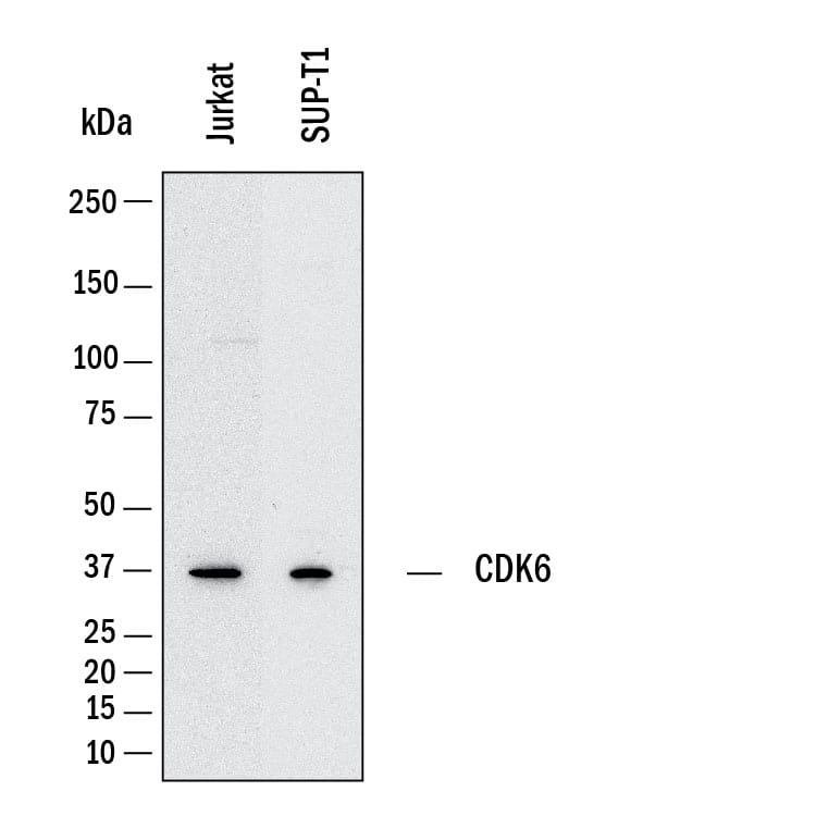
Detection of Human Cdk6 by Western Blot. Western Blot shows lysates of Jurkat human acute T cell leukemia cell line and SUP‑T1 human T cell lymphoblastic lymphoma cell line. PVDF membrane was probed with 0.5 µg/ml of Mouse Anti-Human Cdk6 Monoclonal Antibody (Catalog # MAB11636) followed by HRP-conjugated Anti-Mouse IgG Secondary Antibody (Catalog # HAF018). A specific band was detected for Cdk6 at approximately 36 kDa (as indicated). This experiment was conducted under reducing conditions and using Western Blot Buffer Group 1.
 View Larger
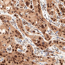
Detection of Cdk6 in Human Lung Cancer. Cdk6 was detected in immersion fixed paraffin-embedded sections of human lung cancer using Mouse Anti-Human Cdk6 Monoclonal Antibody (Catalog # MAB11636) at 5 µg/ml for 1 hour at room temperature followed by incubation with the Anti-Mouse IgG VisUCyte™ HRP Polymer Antibody (Catalog # VC001). Before incubation with the primary antibody, tissue was subjected to heat-induced epitope retrieval using VisUCyte Antigen Retrieval Reagent-Basic (Catalog # VCTS021). Tissue was stained using DAB (brown) and counterstained with hematoxylin (blue). Specific staining was localized to the nucleus and cytoplasm. View our protocol for IHC Staining with VisUCyte HRP Polymer Detection Reagents.
 View Larger
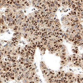
Detection of Cdk6 in Human Pancreatic Cancer. Cdk6 was detected in immersion fixed paraffin-embedded sections of human pancreatic cancer using Mouse Anti-Human Cdk6 Monoclonal Antibody (Catalog # MAB11636) at 5 µg/ml for 1 hour at room temperature followed by incubation with the Anti-Mouse IgG VisUCyte™ HRP Polymer Antibody (Catalog # VC001). Before incubation with the primary antibody, tissue was subjected to heat-induced epitope retrieval using VisUCyte Antigen Retrieval Reagent-Basic (Catalog # VCTS021). Tissue was stained using DAB (brown) and counterstained with hematoxylin (blue). Specific staining was localized to the nucleus and cytoplasm. View our protocol for IHC Staining with VisUCyte HRP Polymer Detection Reagents.
 View Larger
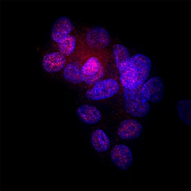
Detection of Cdk6 in HepG2 Human Cell Line. Cdk6 was detected in immersion fixed HepG2 human hepatocellular carcinoma cell line using Mouse Anti-Human Cdk6 Monoclonal Antibody (Catalog # MAB11636) at 8 µg/ml for 3 hours at room temperature. Cells were stained using the NorthernLights™ 557-conjugated Anti-Mouse IgG Secondary Antibody (red; Catalog # NL007) and counterstained with DAPI (blue). Specific staining was localized to the nucleus and cytoplasm. View our protocol for Fluorescent ICC Staining of Cells on Coverslips.
 View Larger
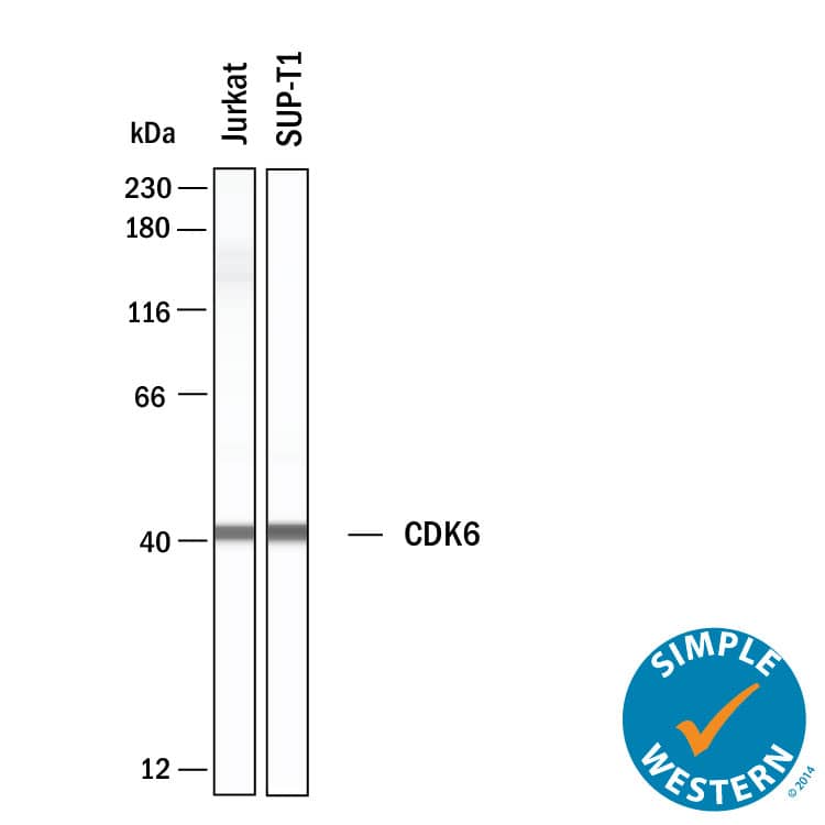
Detection of Human Cdk6 by Simple WesternTM. Simple Western shows lysates of Jurkat human acute T cell leukemia cell line and SUP‑T1 human T cell lymphoblastic lymphoma cell line, loaded at 0.5 mg/ml. A specific band was detected for Cdk6 at approximately 41 kDa (as indicated) using 10 µg/mL of Mouse Anti-Human Cdk6 Monoclonal Antibody (Catalog # MAB11636). This experiment was conducted under reducing conditions and using the 12-230 kDa separation system.
Preparation and Storage
- 12 months from date of receipt, -20 to -70 °C as supplied.
- 1 month, 2 to 8 °C under sterile conditions after reconstitution.
- 6 months, -20 to -70 °C under sterile conditions after reconstitution.
Background: Cdk6
Cell division protein kinase 6 (CDK6) is an enzyme that is regulated by Cyclin D proteins and Cyclin-dependent kinase inhibitor proteins. CDK6 plays an important role in hematopoiesis and loss of CDK6 causes mild anemia, thymic atrophy and delayed G1 progression in lymphocytes. Overexpression of CDK6 has been reported in T-cell lymphoblastic lymphoma and leukemia and in all B-lymphoid malignancies.
- Meyerson M, Harlow E. Identification of G1 kinase activity for cdk6, a novel cyclin D partner. Mol Cell Biol. 1994 Mar;14(3):2077-86. doi: 10.1128/mcb.14.3.2077-2086.1994. PMID: 8114739; PMCID: PMC358568.
- Nebenfuehr S, Kollmann K, Sexl V. The role of CDK6 in cancer. Int J Cancer. 2020 Dec 1;147(11):2988-2995. doi: 10.1002/ijc.33054. Epub 2020 May 30. PMID: 32406095; PMCID: PMC7586846.
Product Datasheets
FAQs
No product specific FAQs exist for this product, however you may
View all Antibody FAQsImmunohistochemistry Reagents
Isotype Controls
Reconstitution Buffers
Secondary Antibodies
Reviews for Human Cdk6 Antibody
There are currently no reviews for this product. Be the first to review Human Cdk6 Antibody and earn rewards!
Have you used Human Cdk6 Antibody?
Submit a review and receive an Amazon gift card.
$25/€18/£15/$25CAN/¥75 Yuan/¥2500 Yen for a review with an image
$10/€7/£6/$10 CAD/¥70 Yuan/¥1110 Yen for a review without an image

