Human CREG Antibody Summary
Arg32-Gln220
Accession # O75629
Customers also Viewed
Applications
Please Note: Optimal dilutions should be determined by each laboratory for each application. General Protocols are available in the Technical Information section on our website.
Scientific Data
 View Larger
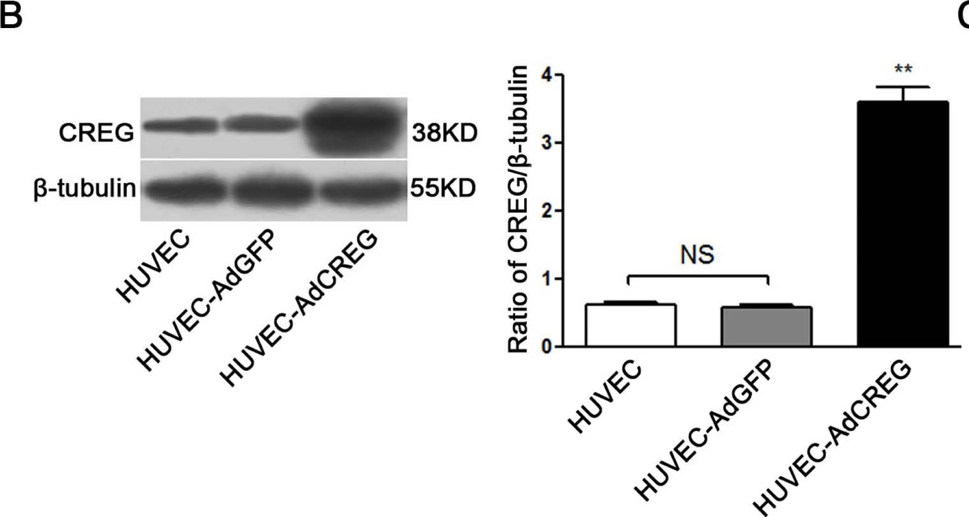
Detection of Human CREG by Western Blot Overexpression of cellular repressor of E1A-stimulated genes (CREG) promotes the proliferation of human umbilical vein endothelial cells (HUVEC). (A) HUVECs infected with adenoviruses carrying CREG-IRES-GFP or GFP, respectively, have a similar infective efficiency. Images were taken by phase contrast microscope (a,c) and by fluorescence microscope (b,d) 48 h after infection. Bar = 25 μm; (B) CREG expression was detected by Western blotting. Levels of CREG were assessed after normalization to beta -tubulin. Data are given as the mean ± SD (n = 3). **p < 0.01 compared with uninfected HUVEC and HUVEC-expressing GFP (AdGFP) control groups. NS: no significant difference; (C) Growth curves of HUVECs, HUVEC-overexpressing CREG (AdCREG) and HUVEC-AdGFP were constructed by plotting cell numbers counted by hemocytometer over three days of incubation. **p < 0.01 compared with uninfected HUVEC and HUVEC-AdGFP control groups. Data are given as the mean ± SD (n = 3). (D) Flow cytometry (FCM) analysis of the cell cycle distributions of cells in three groups. Levels of proliferative potential were accessed by the percentage of cells in the (S + G2) phases of the cell cycle. ***p < 0.001 compared with HUVEC and HUVEC-AdGFP groups. NS: no significant difference. Data are given as the mean ± SD (n = 3). (E) 5-bromo-2′-deoxy-uridine (BrdU) incorporation assay of cells stained positively by diaminobenzidine (DAB) staining solution. The upper panel is a four-times high magnification image of the framed part in the lower panel. Levels of proliferation were assessed by the ratio of average BrdU positive cells to total cells in five random high-magnification fields. Data are given as the mean ± SD (n = 3). * p < 0.05 compared with HUVEC and HUVEC-AdGFP groups. NS: no significant difference. Bar = 25 μm. Image collected and cropped by CiteAb from the following open publication (https://pubmed.ncbi.nlm.nih.gov/24018888), licensed under a CC-BY license. Not internally tested by R&D Systems.
 View Larger
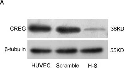
Detection of Human CREG by Western Blot Downregulation of CREG suppresses the proliferation of HUVEC. (A) Stable HUVEC clones with CREG silenced down (H–S) or expressing a scrambled negative control shRNA sequence (scramble) were established by retroviral infection and puromycin selection. Cell lysates were collected, and the expression of CREG was detected by Western blotting with beta -tubulin used as a loading control; (B) Pooled analysis of CREG protein levels assessed after normalization to CREG/ beta -tubulin of the HUVEC group. Scramble HUVEC were used as a negative control. Data are given as the mean ± SD (n = 3). *p < 0.05; ***p < 0.001; NS: no significant difference; (C) Cell counting by hemocytometer. A total of 2 × 10⁵ cells in the logarithmic phase in each of the three groups were plated in four 10-cm diameter dishes. After 24 h, cells were trypsinized and counted by hemocytometer. **p < 0.01 compared with the HUVEC and scramble group; (D) BrdU incorporation assay. Data are given as the mean ± SD (n = 3). **p < 0.01; ***p < 0.001; NS: no significant difference; (E) Effects of the downregulation of CREG on HUVEC cell cycle progression assessed by FCM; (F) Cell proliferation was accessed by the percentage of (S + G2) phase cells in the cell cycle. Data are given as the mean ± SD (n = 3). **p < 0.01 compared with the HUVEC and scramble group. NS: no significant difference. Image collected and cropped by CiteAb from the following open publication (https://pubmed.ncbi.nlm.nih.gov/24018888), licensed under a CC-BY license. Not internally tested by R&D Systems.
Preparation and Storage
- 12 months from date of receipt, -20 to -70 °C as supplied.
- 1 month, 2 to 8 °C under sterile conditions after reconstitution.
- 6 months, -20 to -70 °C under sterile conditions after reconstitution.
Background: CREG
CREG is a secreted glycoprotein that belongs to the CREG protein family. It is induced during differentiation of pluripotent cells and is ubiquitously expressed in adult tissues. Mannose-6 phosphate (M6P) modified CREG binds directly to the M6P/IGF2 receptor to inhibit cell growth (1). Human and mouse CREG share approximately 77% amino acid sequence homology.
Product Datasheets
Citation for Human CREG Antibody
R&D Systems personnel manually curate a database that contains references using R&D Systems products. The data collected includes not only links to publications in PubMed, but also provides information about sample types, species, and experimental conditions.
1 Citation: Showing 1 - 1
-
Cellular repressor of E1A-stimulated genes inhibits human vascular smooth muscle cell apoptosis via blocking P38/JNK MAP kinase activation.
Authors: Han Y, Wu G, Deng J, Tao J, Guo L, Tian X, Kang J, Zhang X, Yan C
J. Mol. Cell. Cardiol., 2010-01-06;48(6):1225-35.
Species: Human
Sample Types: Cell Lysates, Whole Tissue
Applications: IHC-Fr, Western Blot
FAQs
No product specific FAQs exist for this product, however you may
View all Antibody FAQsIsotype Controls
Reconstitution Buffers
Secondary Antibodies
Reviews for Human CREG Antibody
There are currently no reviews for this product. Be the first to review Human CREG Antibody and earn rewards!
Have you used Human CREG Antibody?
Submit a review and receive an Amazon gift card.
$25/€18/£15/$25CAN/¥75 Yuan/¥2500 Yen for a review with an image
$10/€7/£6/$10 CAD/¥70 Yuan/¥1110 Yen for a review without an image


