Human Cytokeratin 18 Antibody Summary
Ala239-Asp397
Accession # P05783
Customers also Viewed
Applications
Please Note: Optimal dilutions should be determined by each laboratory for each application. General Protocols are available in the Technical Information section on our website.
Scientific Data
 View Larger
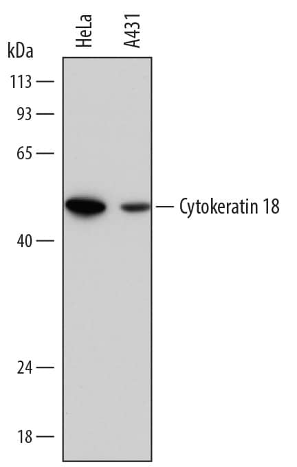
Detection of Human Cytokeratin 18 by Western Blot. Western blot shows lysates of HeLa human cervical epithelial carcinoma cell line and A431 human epithelial carcinoma cell line. PVDF membrane was probed with 0.1 µg/mL of Sheep Anti-Human Cytokeratin 18 Antigen Affinity-purified Polyclonal Antibody (Catalog # AF7619) followed by HRP-conjugated Anti-Sheep IgG Secondary Antibody (HAF016). A specific band was detected for Cytokeratin 18 at approximately 46 kDa (as indicated). This experiment was conducted under reducing conditions and using Immunoblot Buffer Group 1.
 View Larger
Detection of Human Cytokeratin 18 by Western Blot. Western blot shows lysates of HeLa human cervical epithelial carcinoma cell line (Cytokeratin 18 positive) and MOLT‑4 human acute lymphoblastic leukemia cell line (Cytokeratin 18 negative). PVDF membrane was probed with 1 µg/mL of Sheep Anti-Human Cytokeratin 18 Antigen Affinity-purified Polyclonal Antibody (Catalog # AF7619) followed by HRP-conjugated Anti-Sheep IgG Secondary Antibody (HAF016). A specific band was detected for Cytokeratin 18 at approximately 45 kDa (as indicated). GAPDH (AF5718) is shown as a loading control. This experiment was conducted under reducing conditions and using Western Blot Buffer Group 1.
 View Larger
Cytokeratin 18 in HeLa Human Cell Line. Cytokeratin 18 was detected in immersion fixed HeLa human cervical epithelial carcinoma cell line using Sheep Anti-Human Cytokeratin 18 Antigen Affinity-purified Polyclonal Antibody (Catalog # AF7619) at 5 µg/mL for 3 hours at room temperature. Cells were stained using the NorthernLights™ 557-conjugated Anti-Sheep IgG Secondary Antibody (red; NL010) and counterstained with DAPI (blue). Specific staining was localized to cytoskeletal fibers. View our protocol for Fluorescent ICC Staining of Cells on Coverslips.
 View Larger
Cytokeratin 18 in Human Breast Cancer Tissue. Cytokeratin 18 was detected in immersion fixed paraffin-embedded sections of human breast cancer tissue using Sheep Anti-Human Cytokeratin 18 Antigen Affinity-purified Polyclonal Antibody (Catalog # AF7619) at 3 µg/mL overnight at 4 °C. Before incubation with the primary antibody, tissue was subjected to heat-induced epitope retrieval using Antigen Retrieval Reagent-Basic (CTS013). Tissue was stained using the Anti-Sheep HRP-DAB Cell & Tissue Staining Kit (brown; Catalog # CTS019) and counterstained with hematoxylin (blue). Specific staining was localized to intermediate filaments. View our protocol for Chromogenic IHC Staining of Paraffin-embedded Tissue Sections.
 View Larger
Detection of Human Cytokeratin 18 by Simple WesternTM. Simple Western lane view shows lysates of HeLa human cervical epithelial carcinoma cell line and A431 human epithelial carcinoma cell line, loaded at 0.2 mg/mL. A specific band was detected for Cytokeratin 18 at approximately 57 kDa (as indicated) using 1 µg/mL of Sheep Anti-Human Cytokeratin 18 Antigen Affinity-purified Polyclonal Antibody (Catalog # AF7619) followed by 1:50 dilution of HRP-conjugated Anti-Sheep IgG Secondary Antibody (HAF016). This experiment was conducted under reducing conditions and using the 12-230 kDa separation system.
 View Larger
Western Blot Shows Human Cytokeratin 18 Specificity by Using Knockout Cell Line. Western blot shows lysates of HeLa human cervical epithelial carcinoma cell line and human KRT-18 knockout HeLa human cervical epithelial carcinoma cell line (KO). PVDF membrane was probed with 1 µg/mL of Sheep Anti-Human Cytokeratin 18 Antigen Affinity-purified Polyclonal Antibody (Catalog # AF7619) followed by HRP-conjugated Anti-Sheep IgG Secondary Antibody (HAF016). A specific band was detected for Cytokeratin 18 at approximately 48 kDa (as indicated) in the parental HeLa human cervical epithelial carcinoma cell line, but is not detectable in knockout HeLa human cervical epithelial carcinoma cell line. GAPDH (AF5718) is shown as a loading control. This experiment was conducted under reducing conditions and using Western Blot Buffer Group 1.
 View Larger
Detection of Cytokeratin 18 in HeLa Cells (Positive) and HeLa KRT18 Knockout (Negative) Control. Cytokeratin 18 was detected in immersion fixed HeLa Human Cervical Epithelial Carcinoma Cells (Positive) and absent in HeLa KRT18 Knockout (Negative) Control using Sheep Anti-Human Cytokeratin 18 Antigen Affinity-purified Polyclonal Antibody (Catalog # AF7619) at 5 µg/mL for 3 hours at room temperature. Cells were stained using the NorthernLights™ 557-conjugated Anti-Sheep IgG Secondary Antibody (red; Catalog # NL010) and counterstained with DAPI (blue). Specific staining was localized to cytoplasm. View our protocol for Fluorescent ICC Staining of Cells on Coverslips.
Preparation and Storage
- 12 months from date of receipt, -20 to -70 °C as supplied.
- 1 month, 2 to 8 °C under sterile conditions after reconstitution.
- 6 months, -20 to -70 °C under sterile conditions after reconstitution.
Background: Cytokeratin 18
Cytokeratin 18; also KRT-18 (Keratin, type I cytoskeletal 18), Cell proliferation-inducing gene 46 and Keratin-18) is a 44-46 kDa Class I (large keratins of acidic pH) member of the intermediate filament family of proteins. Individual keratins are always expressed in tandem with a second keratin, and these are found in all epithelial cells. The class I Cytokeratin 18 heterodimerizes/polymerizes with 50-52 kDa class II KRT-8 to form 8-10 nm filaments in single strata plus hepatic epithelia. Cytokeratin 18 and -8 are the first keratins to appear in the mammalian embyro. In the adult, Cytokeratin 18 appears to participate in subtractions and additions to the plasma membrane. In this regard, a number of intracellular proteins interact with Cytokeratin 18, including 14-3-3, HSPc70 and Mrj. Cytokeratin 18 may also be O‑glycosylated, and when so, serves to promote Akt-1 activity, thus protecting against apoptosis. Human Cytokeratin 18 is 430 amino acids (aa) in length. It contains an N-terminal "head" region (aa 1-79), a subsequent "rod" region (aa 80-387) with two coiled segments, and a C-terminal tail region. Cytokeratin 18 possesses at least 19 utilized phosphorylation sites plus five acetylated Lys residues. There are multiple isoforms that range from 20-40 kDa in size and are the result of caspase cleavage. A principal cleavage site occurs after Asp238. Over aa 239-397, human Cytokeratin 18 shares 86% aa sequence identity with mouse Cytokeratin 18.
Product Datasheets
Citations for Human Cytokeratin 18 Antibody
R&D Systems personnel manually curate a database that contains references using R&D Systems products. The data collected includes not only links to publications in PubMed, but also provides information about sample types, species, and experimental conditions.
2
Citations: Showing 1 - 2
Filter your results:
Filter by:
-
TFAP2C- and p63-Dependent Networks Sequentially Rearrange Chromatin Landscapes to Drive Human Epidermal Lineage Commitment
Authors: L Li, Y Wang, JL Torkelson, G Shankar, JM Pattison, HH Zhen, F Fang, Z Duren, J Xin, S Gaddam, SP Melo, SN Piekos, J Li, EJ Liaw, L Chen, R Li, M Wernig, WH Wong, HY Chang, AE Oro
Cell Stem Cell, 2019-01-24;0(0):.
Species: Human
Sample Types: Whole Cells
Applications: ICC -
Retinoic acid and BMP4 cooperate with p63 to alter chromatin dynamics during surface epithelial commitment
Authors: JM Pattison, SP Melo, SN Piekos, JL Torkelson, E Bashkirova, MR Mumbach, C Rajasingh, HH Zhen, L Li, E Liaw, D Alber, AJ Rubin, G Shankar, X Bao, HY Chang, PA Khavari, AE Oro
Nat. Genet., 2018-11-05;0(0):.
Species: Human
Sample Types: Whole Cells
Applications: ICC
FAQs
No product specific FAQs exist for this product, however you may
View all Antibody FAQsIsotype Controls
Reconstitution Buffers
Secondary Antibodies
Reviews for Human Cytokeratin 18 Antibody
There are currently no reviews for this product. Be the first to review Human Cytokeratin 18 Antibody and earn rewards!
Have you used Human Cytokeratin 18 Antibody?
Submit a review and receive an Amazon gift card.
$25/€18/£15/$25CAN/¥75 Yuan/¥2500 Yen for a review with an image
$10/€7/£6/$10 CAD/¥70 Yuan/¥1110 Yen for a review without an image


