Human FAK Antibody Summary
Asp213-Thr412
Accession # Q05397
Customers also Viewed
Applications
Please Note: Optimal dilutions should be determined by each laboratory for each application. General Protocols are available in the Technical Information section on our website.
Scientific Data
 View Larger
Detection of Human FAK by Western Blot. Western blot shows lysates of MCF-7 human breast cancer cell line, A549 human lung carcinoma cell line, Huh-7 human hepatoma cell line, and HUVEC human umbilical vein endothelial cells. PVDF membrane was probed with 2 µg/mL of Human FAK Monoclonal Antibody (Catalog # MAB4467) followed by HRP-conjugated Anti-Mouse IgG Secondary Antibody (HAF007). A specific band was detected for FAK at approximately 125 kDa (as indicated). This experiment was conducted under reducing conditions and using Immunoblot Buffer Group 3.
 View Larger
FAK in Human Brain. FAK was detected in immersion fixed paraffin-embedded sections of human brain (hippocampus) using Human FAK Monoclonal Antibody (Catalog # MAB4467) at 25 µg/mL overnight at 4 °C. Tissue was stained using the Anti-Mouse HRP-DAB Cell & Tissue Staining Kit (brown; Catalog # CTS002) and counterstained with hematoxylin (blue). View our protocol for Chromogenic IHC Staining of Paraffin-embedded Tissue Sections.
 View Larger
Western Blot Shows Human FAK Specificity by Using Knockout Cell Line. Western blot shows lysates of HEK293T human embryonic kidney parental cell line and FAK knockout HEK293T cell line (KO). PVDF membrane was probed with 2 µg/mL of Mouse Anti-Human FAK Monoclonal Antibody (Catalog # MAB4467) followed by HRP-conjugated Anti-Mouse IgG Secondary Antibody (HAF018). A specific band was detected for FAK at approximately 135 kDa (as indicated) in the parental HEK293T cell line, but is not detectable in knockout HEK293T cell line. GAPDH (MAB5718) is shown as a loading control. This experiment was conducted under reducing conditions and using Immunoblot Buffer Group 1.
 View Larger
Detection of Human FAK by Simple WesternTM. Simple Western lane view shows lysates of MCF‑7 human breast cancer cell line and A549 human lung carcinoma cell line, loaded at 0.2 mg/mL. A specific band was detected for FAK at approximately 120 kDa (as indicated) using 20 µg/mL of Mouse Anti-Human FAK Monoclonal Antibody (Catalog # MAB4467). This experiment was conducted under reducing conditions and using the 12-230 kDa separation system.
 View Larger
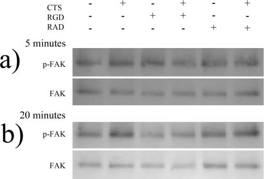
Detection of Human FAK by Western Blot Phosphorylation of FAK following treatment of AF cells derived from non-degenerate IVDs with 1.0 Hz CTS, with and without pre-treatment with RGD or RAD peptides.AF cells derived from non-degenerate IVDs (n = 4) were treated+/− RGD (50 µg/ml) or RAD (50 µg/ml) peptides, and mechanically stimulated (10% CTS, 1.0 Hz frequency) in serum-free media and total protein extracted at timepoints of 5 and 20 minutes. Mechanically stimulated and unstimulated,+/− RGD or RAD peptides, non-degenerate protein samples (5 µg/well) exposed to A) 5 minutes and B) 20 minutes of CTS, were separated using 10% SDS-PAGE and probed using primary antibodies against phosphorylated FAK. Blots were then stripped using a stripping buffer, re-blocked and probed using an antibody against total FAK protein. C) The density of bands were quantified using a Syngene imaging system and the ratio of phosphorylated: total FAK protein normalised to timepoint controls and plotted as % change. *denotes a significant change (p≤0.05) between treatment groups. Image collected and cropped by CiteAb from the following open publication (https://pubmed.ncbi.nlm.nih.gov/24039840), licensed under a CC-BY license. Not internally tested by R&D Systems.
Preparation and Storage
- 12 months from date of receipt, -20 to -70 °C as supplied.
- 1 month, 2 to 8 °C under sterile conditions after reconstitution.
- 6 months, -20 to -70 °C under sterile conditions after reconstitution.
Background: FAK
Focal adhesion kinase 1 (FAK), also known as FAK1 and PTK2, is a ubiquitously expressed non-receptor protein tyrosine kinase that is concentrated in focal adhesions. This cellular localization is directed by a C-terminal 125 amino acid "Focal Adhesion Targeting" (FAT) sequence. FAK plays an important role in migration, cell spreading, differentiation and apoptosis. It associates with several different signaling proteins, such as Src-family PTKs, p130Cas, Shc, Grb2, PI 3-kinase, and Paxillin. These associations enable FAK to function within a network of integrin-stimulated signaling pathways, leading to the activation of targets such as the ERK and JNK mitogen-activated protein kinase pathways. Increased expression and/or activity of FAK in various cancers has been correlated with enhanced proliferation, migration and invasiveness of human tumor cells.
Product Datasheets
Citations for Human FAK Antibody
R&D Systems personnel manually curate a database that contains references using R&D Systems products. The data collected includes not only links to publications in PubMed, but also provides information about sample types, species, and experimental conditions.
2
Citations: Showing 1 - 2
Filter your results:
Filter by:
-
A Three-Dimensional Xeno-Free Culture Condition for Wharton's Jelly-Mesenchymal Stem Cells: The Pros and Cons
Authors: B Koh, N Sulaiman, MB Fauzi, JX Law, MH Ng, TL Yuan, AGN Azurah, MH Mohd Yunus, RBH Idrus, MD Yazid
International Journal of Molecular Sciences, 2023-02-13;24(4):.
Species: Human
Sample Types: Cell Lysates
Applications: Simple Western -
Integrin – Dependent Mechanotransduction in Mechanically Stimulated Human Annulus Fibrosus Cells: Evidence for an Alternative Mechanotransduction Pathway Operating with Degeneration
Authors: Hamish T. J. Gilbert, Navraj S. Nagra, Anthony J. Freemont, Sarah J. Millward-Sadler, Judith A. Hoyland
PLoS ONE
FAQs
No product specific FAQs exist for this product, however you may
View all Antibody FAQsIsotype Controls
Reconstitution Buffers
Secondary Antibodies
Reviews for Human FAK Antibody
There are currently no reviews for this product. Be the first to review Human FAK Antibody and earn rewards!
Have you used Human FAK Antibody?
Submit a review and receive an Amazon gift card.
$25/€18/£15/$25CAN/¥75 Yuan/¥2500 Yen for a review with an image
$10/€7/£6/$10 CAD/¥70 Yuan/¥1110 Yen for a review without an image


