Human IL-1 RII Antibody Summary
Phe14-Glu343 (Glu297Gly)
Accession # P27930
*Small pack size (-SP) is supplied either lyophilized or as a 0.2 µm filtered solution in PBS.
Customers also Viewed
Applications
Please Note: Optimal dilutions should be determined by each laboratory for each application. General Protocols are available in the Technical Information section on our website.
Scientific Data
 View Larger
IL‑1 RII Inhibition of IL‑1 beta /IL‑1F2‑dependent Cell Proliferation and Neutralization by Human IL‑1 RII Antibody. Recombinant Human IL-1 RII (263-2R) inhibits Recombinant Human IL-1 beta / IL-1F2 (201-LB) induced proliferation in the D10.G4.1 mouse helper T cell line in a dose-dependent manner (orange line). Inhibition of Recombinant Human IL-1 beta /IL-1F2 (50 pg/mL) activity elicited by Recombinant Human IL-1 RII (2 µg/mL) is neutralized (green line) by increasing concentrations of Mouse Anti-Human IL-1 RII Monoclonal Antibody (Catalog # MAB263). The ND50 is typically 2-6 µg/mL.
 View Larger
Detection of IL‑1 RII in HDLM-2 (positive) and K562 (negative). IL‑1 RII was detected in immersion fixed HDLM‑2 human Hodgkin’s lymphoma cells (positive) and absent in K562 human chronic myelogenous leukemia cells (negative) using Mouse Anti-Human IL‑1 RII Monoclonal Antibody (Catalog # MAB263) at 25 µg/mL for 3 hours at room temperature. Cells were stained using the NorthernLights™ 557-conjugated Anti-Mouse IgG Secondary Antibody (red; Catalog # NL007) and counterstained with DAPI (blue). Specific staining was localized to cell surface. View our protocol for Fluorescent ICC Staining of Non-adherent Cells.
 View Larger
Detection of IL‑1 RII in HDLM-2 cells by Flow Cytometry HDLM-2 cells were stained with Mouse Anti-Human IL‑1 RII Monoclonal Antibody (Catalog # MAB263, filled histogram) or isotype control antibody (Catalog # MAB003, open histogram) followed by Allophycocyanin-conjugated Anti-Mouse IgG Secondary Antibody (Catalog # F0101B). View our protocol for Staining Membrane-associated Proteins.
 View Larger
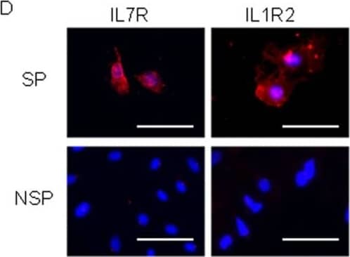
Detection of Human IL-1 RII by Immunocytochemistry/ Immunofluorescence Isolation of SP cells from human first trimester vCTB.(A) Immunocytochemistry images of CGB, CK7, CSH1, HLA−G, CD9 and HLA1 expression in primary vCTB cells isolated by Percoll gradient centrifugation. Each protein expression is shown as red signals (scale bars, 50 µm). The isolated cells were plated on chamber slides with HBM and stained the next day. (B) The SP cells represent 0.12±0.28% (n = 22) of the total primary vCTB. Co-incubation of the cells with verapamil erased the SP cell fraction. (C) The expression of CD105, CD146 and VIM in SP cells from primary vCTB cells was analyzed by immunocytochemistry. All images show counterstaining with Hoechst 33852. Each protein expression is shown as red signals (scale bars, 50 µm). (D) Immunocytochemistry images of IL7R and IL1R2 expression in SP and NSP cells counterstained with Hoechst 33852. IL7R and IL1R2 expressions are shown as red signals (scale bars, 50 µm). (E) The expression of CGB, CSH1 HLA−G, CK7, PHLDA2 and EOMES in IL7R/IL1R2 double-positive cells was assessed by immunocytochemistry. These expressions are shown as green signals (scale bars, 50 µm). (F) Induction of differentiation in IL7R/IL1R2 positive cells was determined by IL7R, IL1R2, CSH1 and CGB expression. IL7R/IL1R2 positive cells from primary human vCTB were analyzed after being cultured in HBM for 2 weeks. IL7R and IL1R2 expressions are shown as red signals; CSH1 and CGB expressions are shown as green signals (scale bars, 50 µm). The cell marked with an arrow show positive for CGB and negative for IL7R expression. In contrast, the cell marked with an arrowhead was positive for IL7R and negative for CGB expression. The outlines of the cells are indicated by broken lines. Image collected and cropped by CiteAb from the following open publication (https://pubmed.ncbi.nlm.nih.gov/21760941), licensed under a CC-BY license. Not internally tested by R&D Systems.
 View Larger
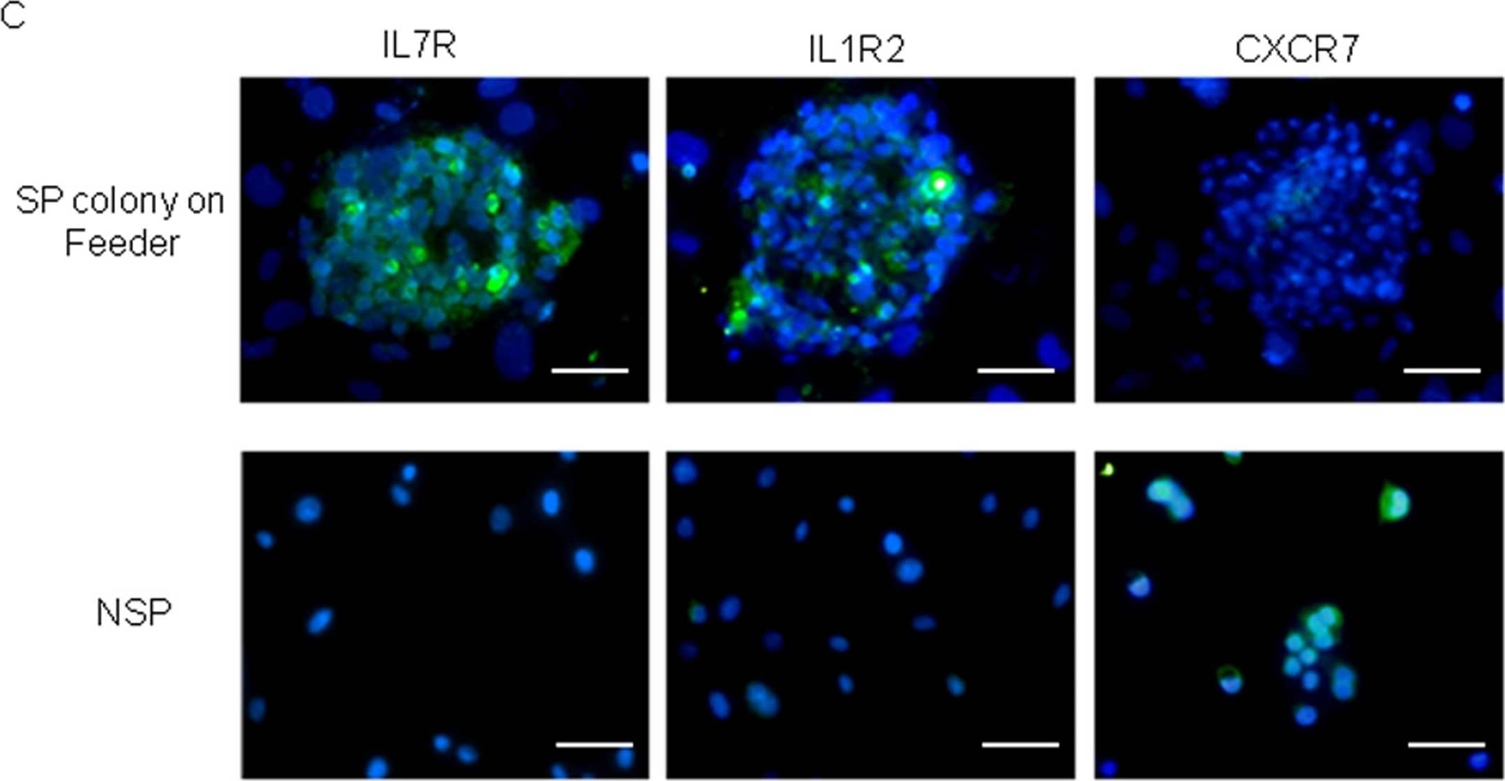
Detection of Human IL-1 RII by Immunocytochemistry/ Immunofluorescence IL7R and IL1R2 were abundantly expressed in HTR-8/SVneo-SP cells.(A) IL7R and IL1R2 mRNA expressions between HTR-8/SVneo-SP and -NSP cells were compared by real-time RT-PCR. The mRNA levels were normalized to those of GAPDH and error bars show standard deviations (n = 3). Asterisk indicates statistically significant (p<0.01). (B) Western blot analysis of IL7R, IL1R2 and CXCR7 proteins in HTR-8/SVneo-SP and -NSP cells. SP cells exclusively expressed IL7R and IL1R2, while NSP exclusively expressed CXCR7. (C) The protein expression of IL7R, IL1R2 or CXCR7 was analyzed by immunocytochemistry. All images counterstained with Hoechst 33852. Each protein expression is shown as green signals (scale bars, 50 µm). (D) IL7R and IL1R2 expressions in HTR-8/SVneo-SP cells were analyzed by immunocytochemistry. All images were counterstained with Hoechst 33852. IL7R and IL1R2 expressions are shown as red and green signals respectively (scale bars, 50 µm). Image collected and cropped by CiteAb from the following open publication (https://pubmed.ncbi.nlm.nih.gov/21760941), licensed under a CC-BY license. Not internally tested by R&D Systems.
 View Larger
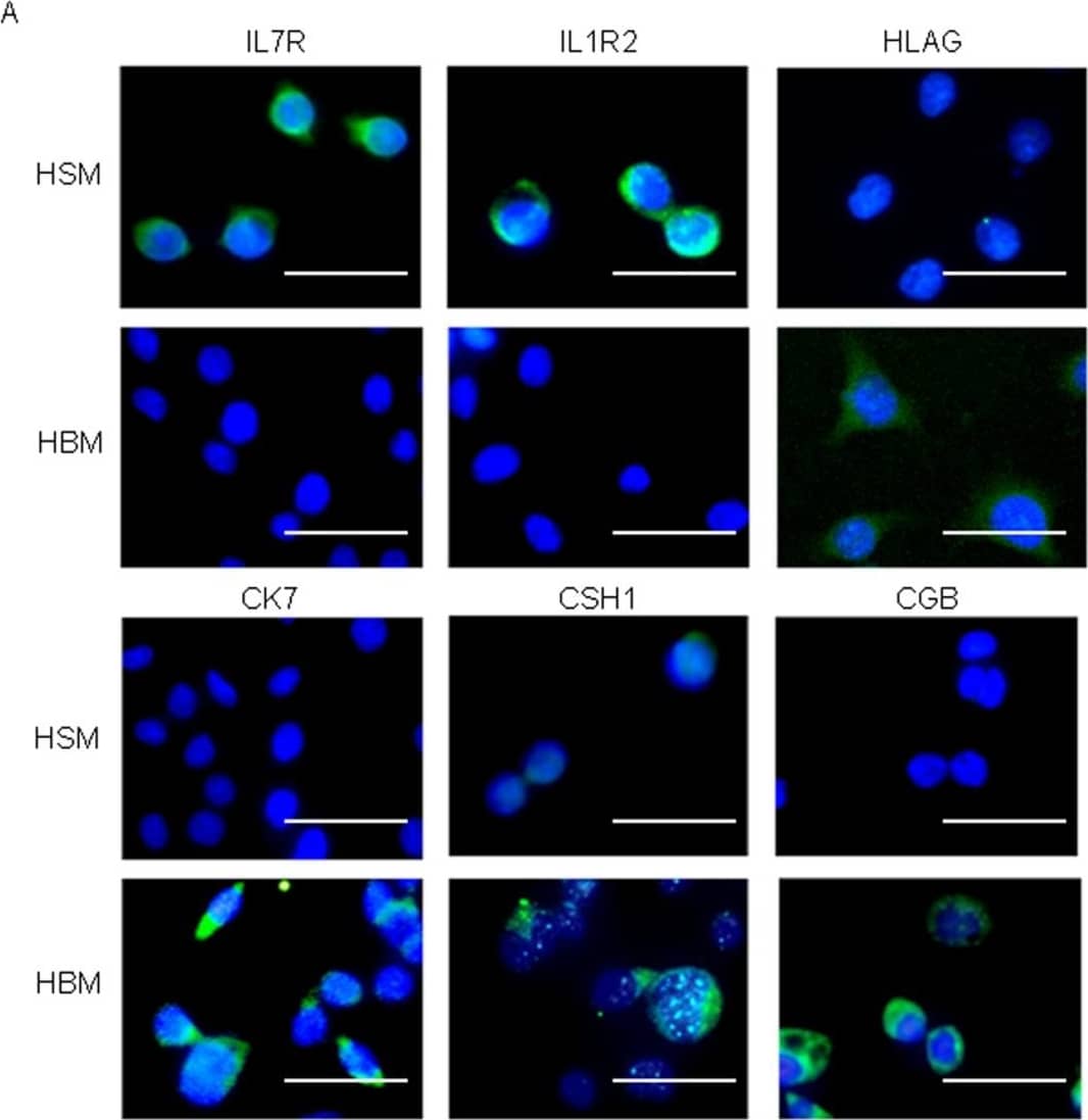
Detection of Human IL-1 RII by Immunocytochemistry/ Immunofluorescence SP and IL1R2/IL7R double-positive cells differentiated into multiple trophoblast cell lineages.(A) Immunocytochemistry images of IL7R, IL1R2, CK7, CSH1, CGB and HLA−G expression in SP cells sorted from HTR-8/SVneo parent cells. Each protein expression is shown as green signals (scale bars, 50 µm). The cells were assayed after culture for 2 weeks in HSM or HBM. (B) Flow cytometry results of IL1R2-positive cells after being sorted by MACS. SP cells were obtained from IL1R2-positive cells after culture for 2 weeks (4.78±1.45% from 3 independent experiments). (C) Induction of trophoblast differentiation of IL7R/IL1R2 double-positive HTR-8/SVneo cells confirmed by differentiation marker, CSH1 and CGB expression. IL7R and IL1R2 expressions are shown as red signals; CSH1 and CGB expressions are shown as green signals (scale bars, 50 µm). The cells marked with an arrow show positive for CSH1 or CGB and negative for IL1R2 or IL7R expression. Image collected and cropped by CiteAb from the following open publication (https://pubmed.ncbi.nlm.nih.gov/21760941), licensed under a CC-BY license. Not internally tested by R&D Systems.
 View Larger
Detection of Human IL-1 RII by Immunocytochemistry/ Immunofluorescence IL7R and IL1R2 were abundantly expressed in HTR-8/SVneo-SP cells.(A) IL7R and IL1R2 mRNA expressions between HTR-8/SVneo-SP and -NSP cells were compared by real-time RT-PCR. The mRNA levels were normalized to those of GAPDH and error bars show standard deviations (n = 3). Asterisk indicates statistically significant (p<0.01). (B) Western blot analysis of IL7R, IL1R2 and CXCR7 proteins in HTR-8/SVneo-SP and -NSP cells. SP cells exclusively expressed IL7R and IL1R2, while NSP exclusively expressed CXCR7. (C) The protein expression of IL7R, IL1R2 or CXCR7 was analyzed by immunocytochemistry. All images counterstained with Hoechst 33852. Each protein expression is shown as green signals (scale bars, 50 µm). (D) IL7R and IL1R2 expressions in HTR-8/SVneo-SP cells were analyzed by immunocytochemistry. All images were counterstained with Hoechst 33852. IL7R and IL1R2 expressions are shown as red and green signals respectively (scale bars, 50 µm). Image collected and cropped by CiteAb from the following open publication (https://pubmed.ncbi.nlm.nih.gov/21760941), licensed under a CC-BY license. Not internally tested by R&D Systems.
Preparation and Storage
- 12 months from date of receipt, -20 to -70 °C as supplied.
- 1 month, 2 to 8 °C under sterile conditions after reconstitution.
- 6 months, -20 to -70 °C under sterile conditions after reconstitution.
Background: IL-1 RII
Two distinct types of receptors that bind the pleiotropic cytokines IL-1 alpha and IL-1 beta have been described. The IL-1 receptor type I is an 80 kDa transmembrane protein that is expressed predominantly by T cells, fibroblasts and endothelial cells. IL-1 receptor type II is a 68 kDa transmembrane protein found on B lymphocytes, neutrophils, monocytes, large granular leukocytes and endothelial cells. Both receptors are members of the immunoglobulin superfamily and show approximately 28% sequence similarity in their extracellular domains. The two receptor types do not heterodimerize in a receptor complex.
An IL-1 receptor accessory protein that can heterodimerize with the type I receptor in the presence of IL-1 alpha or IL-1 beta but not IL-1ra, was identified (1). This type I receptor complex appears to mediate all the known IL-1 biological responses. The receptor type II has a short cytoplasmic domain and does not transduce IL-1 signals. In addition to the membrane-bound form of IL-1 RII, a naturally-occurring soluble form of IL-1 RII has been described. It has been suggested that the type II receptor, either as the membrane-bound or as the soluble form, serves as a decoy for IL-1 and inhibits IL-1 action by blocking the binding of IL-1 to the signaling type I receptor complex.
Product Datasheets
Citations for Human IL-1 RII Antibody
R&D Systems personnel manually curate a database that contains references using R&D Systems products. The data collected includes not only links to publications in PubMed, but also provides information about sample types, species, and experimental conditions.
3
Citations: Showing 1 - 3
Filter your results:
Filter by:
-
Induction of the IL-1RII decoy receptor by NFAT/FOXP3 blocks IL-1 beta -dependent response of Th17 cells
Authors: Dong Hyun Kim, Hee Young Kim, Sunjung Cho, Su-Jin Yoo, Won-Ju Kim, Hye Ran Yeon et al.
eLife
-
Isolation and characterization of human trophoblast side-population (SP) cells in primary villous cytotrophoblasts and HTR-8/SVneo cell line.
Authors: Takao T, Asanoma K, Kato K
PLoS ONE, 2011-07-07;6(7):e21990.
Species: Human
Sample Types: Cell Lysates, Whole Cells
Applications: Cell Selection, ICC, Western Blot -
Immunolocalization of interleukin-1 receptors in the sarcolemma and nuclei of skeletal muscle in patients with idiopathic inflammatory myopathies.
Authors: Grundtman C, Salomonsson S, Dorph C, Bruton J, Andersson U, Lundberg IE
Arthritis Rheum., 2007-02-01;56(2):674-87.
Species: Human
Sample Types: Whole Cells, Whole Tissue
Applications: ICC, IHC-Fr
FAQs
No product specific FAQs exist for this product, however you may
View all Antibody FAQsIsotype Controls
Reconstitution Buffers
Secondary Antibodies
Reviews for Human IL-1 RII Antibody
There are currently no reviews for this product. Be the first to review Human IL-1 RII Antibody and earn rewards!
Have you used Human IL-1 RII Antibody?
Submit a review and receive an Amazon gift card.
$25/€18/£15/$25CAN/¥75 Yuan/¥2500 Yen for a review with an image
$10/€7/£6/$10 CAD/¥70 Yuan/¥1110 Yen for a review without an image


