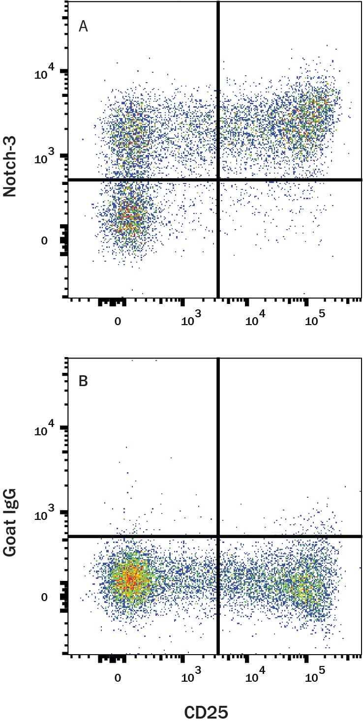
Human Notch-3 Antibody Summary
Ala40-Glu467
Accession # Q9UM47
Customers also Viewed
Applications
Please Note: Optimal dilutions should be determined by each laboratory for each application. General Protocols are available in the Technical Information section on our website.
Preparation and Storage
- 12 months from date of receipt, -20 to -70 °C as supplied.
- 1 month, 2 to 8 °C under sterile conditions after reconstitution.
- 6 months, -20 to -70 °C under sterile conditions after reconstitution.
Background: Notch-3
Human Notch-3 is part of the Notch family of type I transmembrane glycoproteins involved in a number of early-event developmental processes (1). The extracellular domain of Notch receptors interact with the extracellular domain of transmembrane ligands Jagged, Delta, and Serrate expressed on the surface of a neighboring cell. In both vertebrates and invertebrates, Notch signaling is important for specifying cell fates and for defining boundaries between different cell types. The Notch molecule is synthesized as a 2321amino acid (aa) precursor that contains an 39 aa signal sequence, a 1603 aa extracellular region, a 21aa transmembrane (TM) segment and a 658 aa cytoplasmic domain. The large Notch extracellular domain has 34 EGF-like repeats followed by three notch/Lin-12 repeats (LNR) (2). The 11th and 12th EGF-like repeats of Notch have been shown to be both necessary and sufficient for binding the ligands Serrate and Delta, in Drosophila (3). Notch-3 has the same biochemical mechanism of signal tranduction as Notch-1, where a series of cleavage events result in the release of the Notch intracellular domain (NICD). NICD translocates into the nucleus and initiates transcription of Notch-responsive genes (4). Thus, Notch acts as both a ligand-binding receptor and a nuclear factor that regulates transcription.
Mutations in Notch-3 in humans cause an autosomal dominant condition called CADASIL (cerebral autosomal dominant arteriopathy with subcortical infarcts and leukoencephalopathy). This disorder is characterized by recurrent ischemic strokes at an early age without any underlying vascular risk and progressive dementia. Nearly all mutations leading to this disorder are clustered in the first 5 EGF repeats of the Notch-3 gene (5). Human Notch-3 shows 90% aa identity to mouse Notch-3 over the entire protein.
- Weinmaster, G. (2000) Curr. Opin. Genet. Dev. 10:363.
- Joutel, A. et al. (1996) Nature 383:707.
- Rebay, I. et al. (1991) Cell 67:687.
- Mizutani, T. et al. (2001) Proc. Natl. Acad. Sci. USA 98:9026.
- Joutel, A. and E. Tounier-Lasserve (1998) Sem Cell & Dev Biol. 9:619.
Product Datasheets
Citations for Human Notch-3 Antibody
R&D Systems personnel manually curate a database that contains references using R&D Systems products. The data collected includes not only links to publications in PubMed, but also provides information about sample types, species, and experimental conditions.
2
Citations: Showing 1 - 2
Filter your results:
Filter by:
-
Prolyl-isomerase Pin1 controls Notch3 protein expression and regulates T-ALL progression
Oncogene, 2016-02-15;35(36):4741-51.
Species: Human
Sample Types: Whole Cells
Applications: Neutralization -
Notch3 activation modulates cell growth behaviour and cross-talk to Wnt/TCF signalling pathway.
Authors: Wang T, Holt CM, Xu C, Ridley C, P O Jones R, Baron M, Trump D
Cell. Signal., 2007-08-03;19(12):2458-67.
Species: Human
Sample Types: Cell Lysates, Whole Cells
Applications: ICC, Western Blot
FAQs
No product specific FAQs exist for this product, however you may
View all Antibody FAQsIsotype Controls
Reconstitution Buffers
Secondary Antibodies
Reviews for Human Notch-3 Antibody
Average Rating: 4 (Based on 3 Reviews)
Have you used Human Notch-3 Antibody?
Submit a review and receive an Amazon gift card.
$25/€18/£15/$25CAN/¥75 Yuan/¥2500 Yen for a review with an image
$10/€7/£6/$10 CAD/¥70 Yuan/¥1110 Yen for a review without an image
Filter by:


