Human OPA1 Antibody Summary
Accession # O60313
*Small pack size (-SP) is supplied either lyophilized or as a 0.2 µm filtered solution in PBS.
Customers also Viewed


Applications
Please Note: Optimal dilutions should be determined by each laboratory for each application. General Protocols are available in the Technical Information section on our website.
Scientific Data
 View Larger
Detection of Human, Mouse, and Rat OPA1 by Western Blot. Western blot shows lysates of HeLa human cervical epithelial carcinoma cell line, PANC-1 human pancreatic carcinoma cell line, MCF-7 human breast cancer cell line, C2C12 mouse myoblast cell line, and Rat-2 rat embryonic fibroblast cell line. PVDF membrane was probed with 0.2 µg/mL of Rabbit Anti-Human OPA1 Monoclonal Antibody (Catalog # MAB9506) followed by HRP-conjugated Anti-Rabbit IgG Secondary Antibody (Catalog # HAF008). Specific bands were detected for OPA1 at approximately 80-100 kDa (as indicated). This experiment was conducted under reducing conditions and using Immunoblot Buffer Group 1.
 View Larger
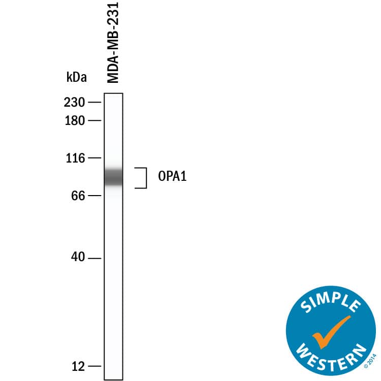
Detection of Human OPA1 by Simple WesternTM. Simple Western shows lysates of MDA‑MB‑231 human breast cancer cell line, loaded at 0.5 mg/ml. A specific band was detected for OPA1 at approximately 90-96 kDa (as indicated) using 20 µg/mL of Rabbit Anti-Human OPA1 Monoclonal Antibody (Catalog # MAB9506). This experiment was conducted under reducing conditions and using the 12-230kDa separation system.
Preparation and Storage
- 12 months from date of receipt, -20 to -70 °C as supplied.
- 1 month, 2 to 8 °C under sterile conditions after reconstitution.
- 6 months, -20 to -70 °C under sterile conditions after reconstitution.
Background: OPA1
Optic Atrophy-1 (OPA1), aka Dynamin-like 120 kDa protein, mitochondrial, is a Dynamin-related GTPase required for mitochondrial fusion and regulation of apoptosis. OPA1 exists as a single-pass membrane protein in the mitochondrion inner membrane as well as in soluble forms in mitochondrion intermembrane space, and is expressed in retina, brain, testis, heart, skeletal muscles. Human OPA1 binds PARL and interacts with CHCHD3 as well as IMMT (preferentially with soluble OPA1 forms). Proteolytic processing in response to intrinsic apoptotic signals may lead to disassembly of OPA1 oligomers and release of the caspase activator cytochrome C (CYCS) into mitochondrial intermembrane space. OPA1 protein form S1 is an inactive form produced by cleavage at S1 position by metalloendopeptidase OMA1 following stress conditions that induce loss of mitochondrial membrane potential, leading to negative regulation of mitochondrial fusion. Defects in OPA1 have been linked to optic atrophy type 1 (OPA1) and dominant optic atrophy plus syndrome (DOA+).
Product Datasheets
FAQs
No product specific FAQs exist for this product, however you may
View all Antibody FAQsIsotype Controls
Reconstitution Buffers
Secondary Antibodies
Reviews for Human OPA1 Antibody
There are currently no reviews for this product. Be the first to review Human OPA1 Antibody and earn rewards!
Have you used Human OPA1 Antibody?
Submit a review and receive an Amazon gift card.
$25/€18/£15/$25CAN/¥75 Yuan/¥2500 Yen for a review with an image
$10/€7/£6/$10 CAD/¥70 Yuan/¥1110 Yen for a review without an image
