Human SLC1A5 Antibody Summary
Accession # Q15758
Customers also Viewed


Applications
Please Note: Optimal dilutions should be determined by each laboratory for each application. General Protocols are available in the Technical Information section on our website.
Scientific Data
 View Larger
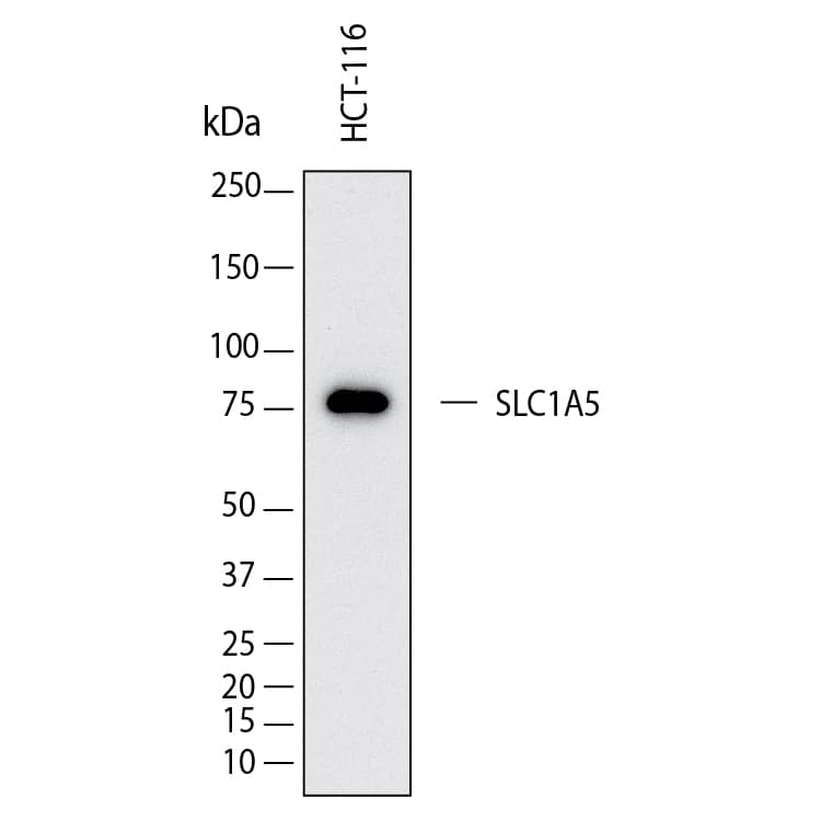
Detection of Human SLC1A5 by Western Blot. Western Blot shows lysates of HCT‑116 human colorectal carcinoma cell line. PVDF membrane was probed with 2 µg/ml of Mouse Anti-Human SLC1A5 Monoclonal Antibody (Catalog # MAB11603) followed by HRP-conjugated Anti-Mouse IgG Secondary Antibody (Catalog # HAF018). A specific band was detected for SLC1A5 at approximately 80 kDa (as indicated). This experiment was conducted under reducing conditions and using Western Blot Buffer Group 1.
 View Larger
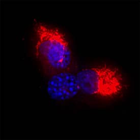
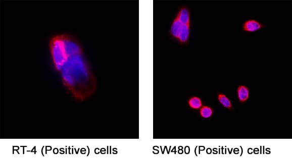
Detection of SLC1A5 in RT-4 cells and SW480 cells. SLC1A5 was detected in immersion fixed RT-4 human bladder carcinoma cell line (Positive) and SW480 human colorectal adenocarcinoma cell line (Positive) using Mouse Anti-Human SLC1A5 Monoclonal Antibody (Catalog # mab11603) at 8 µg/ml for 3 hours at room temperature. Cells were stained using the NorthernLights™ 557-conjugated Anti-Mouse IgG Secondary Antibody (red; Catalog # NL007) and counterstained with DAPI (blue). Specific staining was localized to the cell surface. View our protocol for Fluorescent ICC Staining of Cells on Coverslips.
 View Larger
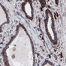
Detection of SLC1A5 in Human Prostate. SLC1A5 was detected in immersion fixed paraffin-embedded sections of human prostate using Mouse Anti-Human SLC1A5 Monoclonal Antibody (Catalog # mab11603) at 0.1 µg/ml for 1 hour at room temperature followed by incubation with the Anti-Mouse IgG VisUCyte™ HRP Polymer Antibody (Catalog # VC001). Before incubation with the primary antibody, tissue was subjected to heat-induced epitope retrieval using VisUCyte Antigen Retrieval Reagent-Basic (Catalog # VCTS021). Tissue was stained using DAB (brown) and counterstained with hematoxylin (blue). Specific staining was localized to the cell membrane of glandular cells. View our protocol for IHC Staining with VisUCyte HRP Polymer Detection Reagents.
Preparation and Storage
- 12 months from date of receipt, -20 to -70 °C as supplied.
- 1 month, 2 to 8 °C under sterile conditions after reconstitution.
- 6 months, -20 to -70 °C under sterile conditions after reconstitution.
Background: SLC1A5
SLC1A5 is a sodium dependent antiporter also known as ASCT2 (ASC amino acid Transporter2). It is a member of the solute carrier (SLC) superfamily of transporters and plays an important role in tumors. It is one of the 3 proteins, including SLC7A5 and SLC3A2, that are among the highest differentially expressed genes in activated lymphocytes and cancerous cells. Pharmacological inhibition of SLC1A5 was found to abrogate the effector functions of NK cells. In a pancancer analysis of SLC1A5, it has been demonstrated to be a prognostic biomarker in cancer patients.
- Nachef M, Ali AK, Almutairi SM, Lee SH. Targeting SLC1A5 and SLC3A2/SLC7A5 as a Potential Strategy to Strengthen Anti-Tumor Immunity in the Tumor Microenvironment. Front Immunol. 2021 Apr 19;12:624324. doi: 10.3389/fimmu.2021.624324. PMID: 33953707; PMCID: PMC8089370.
- Chen P, Jiang Y, Liang J, Cai J, Zhuo Y, Fan H, Yuan R, Cheng S, Zhang Y. SLC1A5 is a novel biomarker associated with ferroptosis and the tumor microenvironment: a pancancer analysis. Aging (Albany NY). 2023 Aug 10;15(15):7451-7475. doi: 10.18632/aging.204911. Epub 2023 Aug 10. PMID: 37566748; PMCID: PMC10457057.
Product Datasheets
FAQs
No product specific FAQs exist for this product, however you may
View all Antibody FAQsImmunohistochemistry Reagents
Isotype Controls
Reconstitution Buffers
Secondary Antibodies
Reviews for Human SLC1A5 Antibody
There are currently no reviews for this product. Be the first to review Human SLC1A5 Antibody and earn rewards!
Have you used Human SLC1A5 Antibody?
Submit a review and receive an Amazon gift card.
$25/€18/£15/$25CAN/¥75 Yuan/¥2500 Yen for a review with an image
$10/€7/£6/$10 CAD/¥70 Yuan/¥1110 Yen for a review without an image

