Mouse B7-H3 Antibody Summary
Val29-Phe244
Accession # Q8VE98.1
Customers also Viewed
Applications
Please Note: Optimal dilutions should be determined by each laboratory for each application. General Protocols are available in the Technical Information section on our website.
Scientific Data
 View Larger
B7‑H3 in NIH‑3T3 Mouse Cell Line. B7‑H3 was detected in immersion fixed NIH‑3T3 mouse embryonic fibroblast cell line (positive staining) and MOLT‑4 human acute lymphoblastic leukemia cell line (negative staining) using Rabbit Anti-Mouse B7‑H3 Monoclonal Antibody (Catalog # MAB10271) at 8 µg/mL for 3 hours at room temperature. Cells were stained using the NorthernLights™ 557-conjugated Anti-Rabbit IgG Secondary Antibody (red; NL004) and counterstained with DAPI (blue). Specific staining was localized to plasma membrane. Staining was performed using our protocol for Fluorescent ICC Staining of Non-adherent Cells.
 View Larger
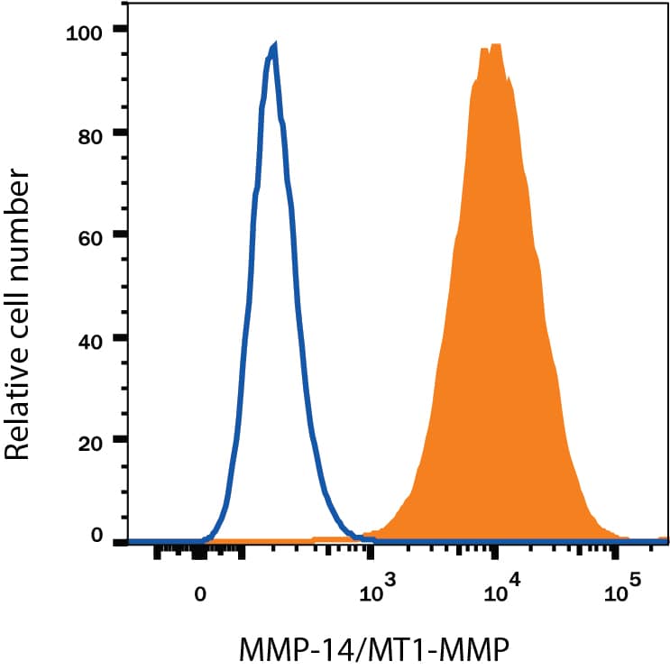
Detection of B7-H3 in C2C12 Mouse Cell Line by Flow Cytometry. C2C12 mouse muscle myoblast cell line was stained with Rabbit Anti-Mouse B7-H3 Monoclonal Antibody (Catalog # MAB10271, filled histogram) or isotype control antibody (MAB1050, open histogram), followed by Phycoerythrin-conjugated Anti-Rabbit IgG F(ab')2Secondary Antibody (F0110).
Preparation and Storage
- 12 months from date of receipt, -20 to -70 °C as supplied.
- 1 month, 2 to 8 °C under sterile conditions after reconstitution.
- 6 months, -20 to -70 °C under sterile conditions after reconstitution.
Background: B7-H3
T cells require a signal induced by the engagement of the T cell receptor and a “costimulatory” signal(s) through distinct T cell surface molecules for optimal T cell expansion and activation. Members of the B7 superfamily of counter-receptors were identified by their ability to interact with costimulatory molecules found on the surface of T cells. Members of the B7 superfamily include B7-1 (CD80), B7-2 (CD86), B7-H1 (PD-L1), B7-H2 (B7RP-1), B7-H3, and PD-L2 (1). B7-H3 is expressed at very high levels in immature dendritic cells at moderate levels on mature dendritic cells, LPS stimulated immature dendritic cells and LPS stimulated monocytes, and at low levels on resting monocytes. B7-H3 binds to activated T cells via an as-of-yet identified receptor. B7-H3 co-stimulates proliferation of T cells and interferon-gamma (IFN-gamma ) production and enhances the induction of cytotoxic T cells. B7-H3 shares 20 - 27% amino acid (aa) identity with other B7 family members (2). Murine B7-H3 is a 259 aa protein containing an extracellular domain, a transmembrane domain and a cytoplasmic domain. Mouse and human B7-H3 share 87% aa identity (3).
- Coyle, A.J. and J.-C. Gutierrez-Ramos (2001) Nature Immunol. 2:203.
- Chapoval, A.I. et al. (2001) Nature Immunol. 2:269.
- Sun, M. et al. (2002) J. Immunol. 168:6294.
Product Datasheets
FAQs
No product specific FAQs exist for this product, however you may
View all Antibody FAQsIsotype Controls
Reconstitution Buffers
Secondary Antibodies
Reviews for Mouse B7-H3 Antibody
There are currently no reviews for this product. Be the first to review Mouse B7-H3 Antibody and earn rewards!
Have you used Mouse B7-H3 Antibody?
Submit a review and receive an Amazon gift card.
$25/€18/£15/$25CAN/¥75 Yuan/¥2500 Yen for a review with an image
$10/€7/£6/$10 CAD/¥70 Yuan/¥1110 Yen for a review without an image


