Human Caspase-1 Antibody Summary
Asn120-Asp297
Accession # P29466
Customers also Viewed
Applications
Please Note: Optimal dilutions should be determined by each laboratory for each application. General Protocols are available in the Technical Information section on our website.
Scientific Data
 View Larger
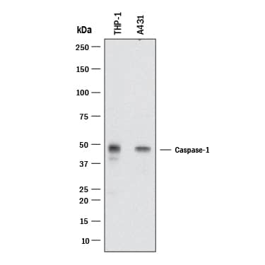
Detection of Human Caspase‑1 by Western Blot. Western blot shows lysates of A431 human epithelial carcinoma cell line and THP-1 human acute monocytic leukemia cell line. PVDF Membrane was probed with 0.25 µg/mL of Goat Anti-Human Caspase-1 Antigen Affinity-purified Polyclonal Antibody (Catalog # AF6215) followed by HRP-conjugated Anti-Goat IgG Secondary Antibody (Catalog # HAF109). A specific band was detected for Caspase-1 at approximately 50 kDa (as indicated). This experiment was conducted under reducing conditions and using Immunoblot Buffer Group 2.
 View Larger
Caspase‑1 in THP‑1 Human Cell Line. Caspase-1 was detected in immersion fixed THP-1 human acute monocytic leukemia cell line using Goat Anti-Human Caspase-1 Antigen Affinity-purified Polyclonal Antibody (Catalog # AF6215) at 15 µg/mL for 3 hours at room temperature. Cells were stained using the NorthernLights™ 557-conjugated Anti-Goat IgG Secondary Antibody (red; Catalog # NL001) and counterstained with DAPI (blue). Specific staining was localized to cytoplasm. View our protocol for Fluorescent ICC Staining of Non-adherent Cells.
 View Larger
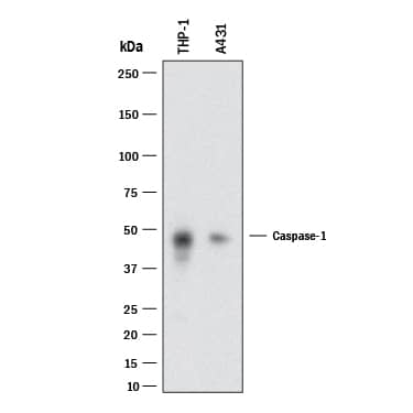
Detection of Human Caspase‑1 by Simple WesternTM. Simple Western lane view shows lysates of A431 human epithelial carcinoma parental cell line and Caspase-1 knockout A431 cell line (KO), loaded at 0.2 mg/mL. A specific band was detected for Caspase-1 at approximately 52 kDa (as indicated) using 25 µg/mL of Goat Anti-Human Caspase-1 Antigen Affinity-purified Polyclonal Antibody (Catalog # AF6215) followed by 1:50 dilution of HRP-conjugated Anti-Goat IgG Secondary Antibody (Catalog # HAF109). This experiment was conducted under reducing conditions and using the 12-230 kDa separation system.
 View Larger
Western Blot Shows Human Caspase‑1 Specificity by Using Knockout Cell Line. Western blot shows lysates of A431 human epithelial carcinoma parental cell line and Caspase-1 knockout A431 cell line (KO). PVDF membrane was probed with 0.5 µg/mL of Goat Anti-Human Caspase-1 Antigen Affinity-purified Polyclonal Antibody (Catalog # AF6215) followed by HRP-conjugated Anti-Goat IgG Secondary Antibody (Catalog # HAF017). A specific band was detected for Caspase-1 at approximately 50 kDa (as indicated) in the parental A431 cell line, but is not detectable in knockout A431 cell line. GAPDH (Catalog # AF5718) is shown as a loading control. This experiment was conducted under reducing conditions and using Immunoblot Buffer Group 1.
Preparation and Storage
- 12 months from date of receipt, -20 to -70 °C as supplied.
- 1 month, 2 to 8 °C under sterile conditions after reconstitution.
- 6 months, -20 to -70 °C under sterile conditions after reconstitution.
Background: Caspase-1
Caspase-1, also known as IL-1 beta -converting enzyme (ICE), is an aspartic protease that plays a key role in the inflammatory response and apoptosis. Caspase-1 precursor (about 50kDa) can be cleaved and the active enzyme consists of a complex of two 20 kDa (aa 120-297) and two 10 kDa (aa 317-404) subunits which associate following cleavage of inactive precursors. Caspase-1 is required for proteolytic cleavage of the IL-1 beta precursor to form the active proinflammatory cytokine. Alternate splicing generates several additional Caspase-1 isoforms with deletions in the propeptide regions or also in the mature subunits. Within the large subunit, human Caspase 1 shares 61% aa sequence identity with mouse and rat Caspase-1.
Product Datasheets
Citations for Human Caspase-1 Antibody
R&D Systems personnel manually curate a database that contains references using R&D Systems products. The data collected includes not only links to publications in PubMed, but also provides information about sample types, species, and experimental conditions.
5
Citations: Showing 1 - 5
Filter your results:
Filter by:
-
NF?B and NLRP3/NLRC4 inflammasomes regulate differentiation, activation and functional properties of monocytes in response to distinct SARS-CoV-2 proteins
Authors: Tsukalov, I;Sánchez-Cerrillo, I;Rajas, O;Avalos, E;Iturricastillo, G;Esparcia, L;Buzón, MJ;Genescà, M;Scagnetti, C;Popova, O;Martin-Cófreces, N;Calvet-Mirabent, M;Marcos-Jimenez, A;Martínez-Fleta, P;Delgado-Arévalo, C;de Los Santos, I;Muñoz-Calleja, C;Calzada, MJ;González Álvaro, I;Palacios-Calvo, J;Alfranca, A;Ancochea, J;Sánchez-Madrid, F;Martin-Gayo, E;
Nature communications
Species: Human
Sample Types: Whole Tissue
Applications: Immunohistochemistry -
A Salmonella Typhi RNA thermosensor regulates virulence factors and innate immune evasion in response to host temperature
Authors: SM Brewer, C Twittenhof, J Kortmann, SW Brubaker, J Honeycutt, LM Massis, THM Pham, F Narberhaus, DM Monack
PloS Pathogens, 2021-03-02;17(3):e1009345.
Species: Human
Sample Types: Cell Lysates
Applications: Western Blot -
Human metapneumovirus activates NOD-like receptor protein 3 inflammasome via its small hydrophobic protein which plays a detrimental role during infection in mice
Authors: VB Lê, J Dubois, C Couture, MH Cavanagh, O Uyar, A Pizzorno, M Rosa-Calat, MÈ Hamelin, G Boivin
PLoS Pathog., 2019-04-09;15(4):e1007689.
Species: Human
Sample Types: Cell Lysates
Applications: Western Blot -
Cutting Edge: Inflammasome Activation in Primary Human Macrophages Is Dependent on Flagellin.
Authors: Kortmann J, Brubaker S, Monack D
J Immunol, 2015-06-24;195(3):815-9.
Species: Human
Sample Types: Cell Culture Supernates
Applications: Western Blot -
Staphylococcus aureus activates the NLRP3 inflammasome in human and rat conjunctival goblet cells.
Authors: McGilligan V, Gregory-Ksander M, Li D, Moore J, Hodges R, Gilmore M, Moore T, Dartt D
PLoS ONE, 2013-09-10;8(9):e74010.
Species: Human, Rat
Sample Types: Cell Lysates
Applications: Western Blot
FAQs
No product specific FAQs exist for this product, however you may
View all Antibody FAQsIsotype Controls
Reconstitution Buffers
Secondary Antibodies
Reviews for Human Caspase-1 Antibody
Average Rating: 4 (Based on 1 Review)
Have you used Human Caspase-1 Antibody?
Submit a review and receive an Amazon gift card.
$25/€18/£15/$25CAN/¥75 Yuan/¥2500 Yen for a review with an image
$10/€7/£6/$10 CAD/¥70 Yuan/¥1110 Yen for a review without an image
Filter by:


