Human M-CSF Antibody Summary
Applications
Human M-CSF Sandwich Immunoassay
Please Note: Optimal dilutions should be determined by each laboratory for each application. General Protocols are available in the Technical Information section on our website.
Preparation and Storage
- 12 months from date of receipt, -20 to -70 °C as supplied.
- 1 month, 2 to 8 °C under sterile conditions after reconstitution.
- 6 months, -20 to -70 °C under sterile conditions after reconstitution.
Background: M-CSF
M-CSF, also known as CSF-1, is a four-alpha-helical-bundle cytokine that is the primary regulator of macrophage survival, proliferation and differentiation. M-CSF is also essential for the survival and proliferation of osteoclast progenitors. M-CSF also primes and enhances macrophage killing of tumor cells and microorganisms, regulates the release of cytokines and other inflammatory modulators from macrophages, and stimulates pinocytosis. M-CSF increases during pregnancy to support implantation and growth of the decidua and placenta. Sources of M-CSF include fibroblasts, activated macrophages, endometrial secretory epithelium, bone marrow stromal cells and activated endothelial cells. The M-CSF receptor (c-fms) transduces its pleotropic effects and mediates its endocytosis. M-CSF mRNAs of various sizes occur. Full length human M-CSF transcripts encode a 522 amino acid (aa) type I transmembrane (TM) protein with a 464 aa extracellular region, a 21 aa TM domain, and a 37 aa cytoplasmic tail that forms a 140 kDa covalent dimer. Differential processing produces two proteolytically cleaved, secreted dimers. One is an N- and O- glycosylated 86 kDa dimer, while the other is modified by both glycosylation and chondroitin-sulfate proteoglycan (PG) to generate a 200 kDa subunit. Although PG-modified M-CSF can circulate, it may be immobilized by attachment to type V collagen. Shorter transcripts encode M-CSF that lack cleavage and PG sites and produce an N-glycosylated 68 kDa TM dimer and a slowly produced 44 kDa secreted dimer. Although forms may vary in activity and half-life, all contain the N-terminal 150 aa portion that is necessary and sufficient for interaction with the M-CSF receptor. The first 223 aa of mature human M-CSF shares 88%, 86%, 81% and 74% aa identity with corresponding regions of dog, cow, mouse and rat M-CSF, respectively. Human M-CSF is active in the mouse, but mouse M-CSF is reported to be species-specific.
Product Datasheets
Citations for Human M-CSF Antibody
R&D Systems personnel manually curate a database that contains references using R&D Systems products. The data collected includes not only links to publications in PubMed, but also provides information about sample types, species, and experimental conditions.
2
Citations: Showing 1 - 2
Filter your results:
Filter by:
-
Factors Influencing the Differentiation of Human Monocytic Myeloid-Derived Suppressor Cells Into Inflammatory Macrophages
Authors: D Bayik, D Tross, DM Klinman
Front Immunol, 2018-03-26;9(0):608.
Species: Human
Sample Types: Cell Culture Supernates
Applications: ELISA Capture -
A multiplex immunoassay for human adipokine profiling.
Authors: Schipper HS, De Jager W, van Dijk ME, Meerding J, Zelissen PM, Adan RA, Prakken BJ, Kalkhoven E
Clin. Chem., 2010-06-08;56(0):1320.
Species: Human
Sample Types: Cell Culture Supernates
Applications: Luminex Development
FAQs
No product specific FAQs exist for this product, however you may
View all Antibody FAQsIsotype Controls
Reconstitution Buffers
Secondary Antibodies
Reviews for Human M-CSF Antibody
Average Rating: 5 (Based on 2 Reviews)
Have you used Human M-CSF Antibody?
Submit a review and receive an Amazon gift card.
$25/€18/£15/$25CAN/¥75 Yuan/¥2500 Yen for a review with an image
$10/€7/£6/$10 CAD/¥70 Yuan/¥1110 Yen for a review without an image
Filter by:
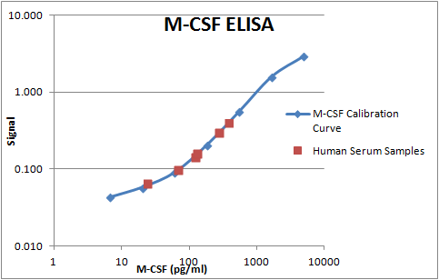
This antibody was used as a capture antibody in the development of a sandwich ELISA. The R&D systems polyclonal antibody AF216 was used as the detection antibody. M-CSF was measured in human serum samples with this assay.


