Human SOX3 Antibody Summary
Met1-Leu230
Accession # P41225
Applications
Please Note: Optimal dilutions should be determined by each laboratory for each application. General Protocols are available in the Technical Information section on our website.
Scientific Data
 View Larger
SOX3 in NTera‑2 Human Cell Line. SOX3 was detected in immersion fixed NTera-2 human testicular embryonic carcinoma cell line differentiation with retinoic acid using Goat Anti-Human SOX3 Antigen Affinity-purified Polyclonal Antibody (Catalog # AF2569) at 10 µg/mL for 3 hours at room temperature. Cells were stained using the NorthernLights™ 493-conjugated Anti-Goat IgG Secondary Antibody (green, upper panel; NL003) and counterstained with DAPI (blue, lower panel). View our protocol for Fluorescent ICC Staining of Cells on Coverslips.
 View Larger
Detection of SOX3 in U‑251 MG cells (positive) and HDLM‑2 cells (negative). SOX3 was detected in immersion fixed U‑251 MG human glioblastoma cells (positive) and absent in HDLM‑2 human Hodgkin’s lymphoma cells (negative) using Goat Anti-Human SOX3 Antigen Affinity-purified Polyclonal Antibody (Catalog # AF2569) at 15 µg/mL for 3 hours at room temperature. Cells were stained using the NorthernLights™ 557-conjugated Anti-Goat IgG Secondary Antibody (red; Catalog # NL001) and counterstained with DAPI (blue). Specific staining was localized to cell nuclei. View our protocol for Fluorescent ICC Staining of Cells on Coverslips.
 View Larger
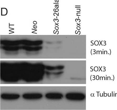
Detection of SOX3 by Western Blot Transcription is unaffected but protein is cleared from mutant cells.A) SOX3 protein is present in every WT cell (NEO−) of the 13.5 dpc telencephalic ventricular zone but virtually absent from equivalent tissue derived from Sox3-26ala cells (NEO+). B) Comparison of SOX3 immunostaining on Sox3-null cells (from a 14.5 dpc +/− embryo) and Sox3-26ala expressing cells (from a Sox3-26ala <-> WT chimera) confirming that the antibody is SOX3-specific and that the Sox3-26ala expressing cells exhibit a low level of residual nuclear protein. C) WT, Neo, Sox3-26ala and Sox3-null ES cells were differentiated for 5 days in CDM as multi-cellular bodies. Rare SOX3 positive cells were detected in Sox3-26ala CDMs while the majority of cells had low SOX3 protein levels in comparison to neighbouring WT CDM bodies processed on the same slide. D–E) WT, Neo, Sox3-26ala and Sox3-null ES cells were grown in N2B27 for 4 days to form neural progenitors. Western blotting for SOX3 reveals a dramatic reduction of protein in Sox3-26ala cells (D); 3 and 30 minute exposures are shown. E) Transcript levels of Sox3 are not affected in Sox3-26ala cells as determined by qPCR. Three experimental replicates are shown. Data was normalised to Sox3 levels inSox3-Neo control cells and error bars represent SEM. F) ISH confirms that Sox3 transcript is present at comparable levels in ventricular zone cells at 13.5 dpc derived from both WT (Neo−) and Sox3-26ala (Neo+) cells. ISH performed on adjacent 10 µm coronal sections of 13.5 dpc chimeric telencephalon. Image collected and cropped by CiteAb from the following open publication (https://pubmed.ncbi.nlm.nih.gov/23505376), licensed under a CC-BY license. Not internally tested by R&D Systems.
 View Larger
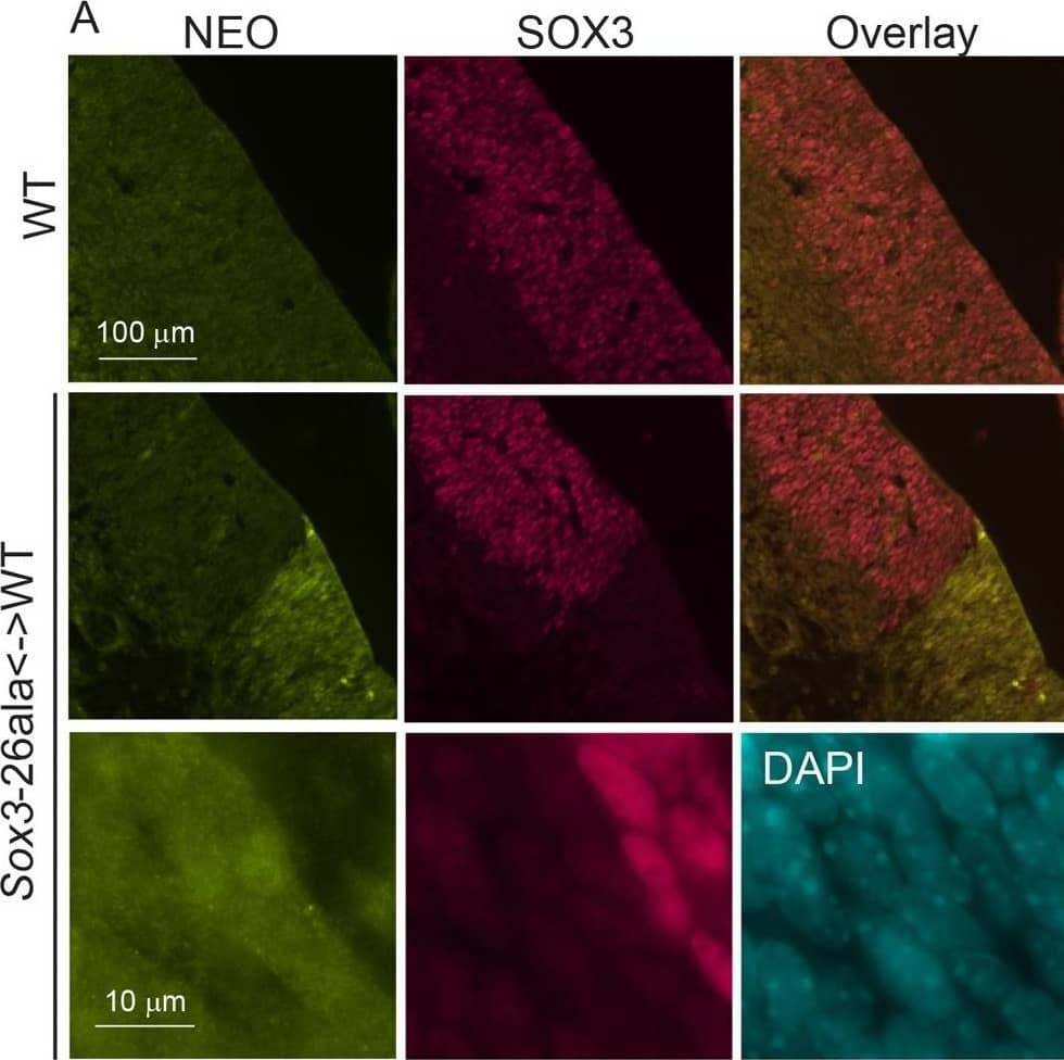
Detection of SOX3 by Immunohistochemistry Transcription is unaffected but protein is cleared from mutant cells.A) SOX3 protein is present in every WT cell (NEO−) of the 13.5 dpc telencephalic ventricular zone but virtually absent from equivalent tissue derived from Sox3-26ala cells (NEO+). B) Comparison of SOX3 immunostaining on Sox3-null cells (from a 14.5 dpc +/− embryo) and Sox3-26ala expressing cells (from a Sox3-26ala <-> WT chimera) confirming that the antibody is SOX3-specific and that the Sox3-26ala expressing cells exhibit a low level of residual nuclear protein. C) WT, Neo, Sox3-26ala and Sox3-null ES cells were differentiated for 5 days in CDM as multi-cellular bodies. Rare SOX3 positive cells were detected in Sox3-26ala CDMs while the majority of cells had low SOX3 protein levels in comparison to neighbouring WT CDM bodies processed on the same slide. D–E) WT, Neo, Sox3-26ala and Sox3-null ES cells were grown in N2B27 for 4 days to form neural progenitors. Western blotting for SOX3 reveals a dramatic reduction of protein in Sox3-26ala cells (D); 3 and 30 minute exposures are shown. E) Transcript levels of Sox3 are not affected in Sox3-26ala cells as determined by qPCR. Three experimental replicates are shown. Data was normalised to Sox3 levels inSox3-Neo control cells and error bars represent SEM. F) ISH confirms that Sox3 transcript is present at comparable levels in ventricular zone cells at 13.5 dpc derived from both WT (Neo−) and Sox3-26ala (Neo+) cells. ISH performed on adjacent 10 µm coronal sections of 13.5 dpc chimeric telencephalon. Image collected and cropped by CiteAb from the following open publication (https://pubmed.ncbi.nlm.nih.gov/23505376), licensed under a CC-BY license. Not internally tested by R&D Systems.
 View Larger
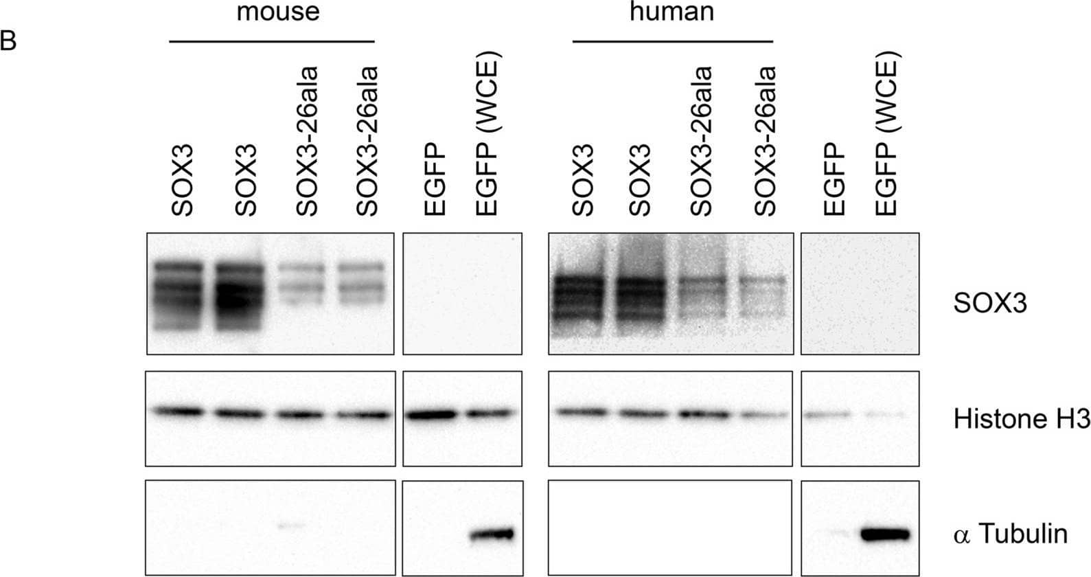
Detection of SOX3 by Western Blot SOX3-26ala from mouse and human retains transactivation activity.A) COS-7 cells were transfected with pcDNA3.1 expression vector containing either mouse Sox3, human SOX3, mouse Sox3-26ala, human SOX3-26ala or an empty vector control. Values represent mean normalised luciferase values plus standard deviation of four independent experiments measured 48 hours after transfection. Student's T-tests (two tailed, unequal variance) of SOX3-26ala from human or mouse compared to empty vector control show a statistically significant increase in luciferase activity. B) Nuclear protein lysates prepared from duplicate plates 48 hours after transfection show that less SOX3 is detected in the nucleus of cells expressing both mouse and human SOX3-26ala. pcDNA3.1-EGFP transfected cells were used as a control and prepared for both nuclear protein and whole cell extracts (WCE). Blotting for Histone H3, indicates equal loading and blotting for alpha -Tubulin shows an absence of cytoplasmic contamination in nuclear preparations. Transfection efficiency was determined by co-transfecting EGFP and counting positive cells prior to harvesting and found to be equal for all plasmids. Image collected and cropped by CiteAb from the following open publication (https://pubmed.ncbi.nlm.nih.gov/23505376), licensed under a CC-BY license. Not internally tested by R&D Systems.
 View Larger
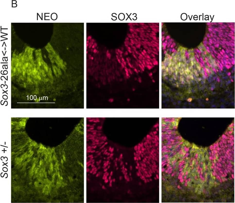
Detection of SOX3 by Immunohistochemistry Transcription is unaffected but protein is cleared from mutant cells.A) SOX3 protein is present in every WT cell (NEO−) of the 13.5 dpc telencephalic ventricular zone but virtually absent from equivalent tissue derived from Sox3-26ala cells (NEO+). B) Comparison of SOX3 immunostaining on Sox3-null cells (from a 14.5 dpc +/− embryo) and Sox3-26ala expressing cells (from a Sox3-26ala <-> WT chimera) confirming that the antibody is SOX3-specific and that the Sox3-26ala expressing cells exhibit a low level of residual nuclear protein. C) WT, Neo, Sox3-26ala and Sox3-null ES cells were differentiated for 5 days in CDM as multi-cellular bodies. Rare SOX3 positive cells were detected in Sox3-26ala CDMs while the majority of cells had low SOX3 protein levels in comparison to neighbouring WT CDM bodies processed on the same slide. D–E) WT, Neo, Sox3-26ala and Sox3-null ES cells were grown in N2B27 for 4 days to form neural progenitors. Western blotting for SOX3 reveals a dramatic reduction of protein in Sox3-26ala cells (D); 3 and 30 minute exposures are shown. E) Transcript levels of Sox3 are not affected in Sox3-26ala cells as determined by qPCR. Three experimental replicates are shown. Data was normalised to Sox3 levels inSox3-Neo control cells and error bars represent SEM. F) ISH confirms that Sox3 transcript is present at comparable levels in ventricular zone cells at 13.5 dpc derived from both WT (Neo−) and Sox3-26ala (Neo+) cells. ISH performed on adjacent 10 µm coronal sections of 13.5 dpc chimeric telencephalon. Image collected and cropped by CiteAb from the following open publication (https://pubmed.ncbi.nlm.nih.gov/23505376), licensed under a CC-BY license. Not internally tested by R&D Systems.
 View Larger
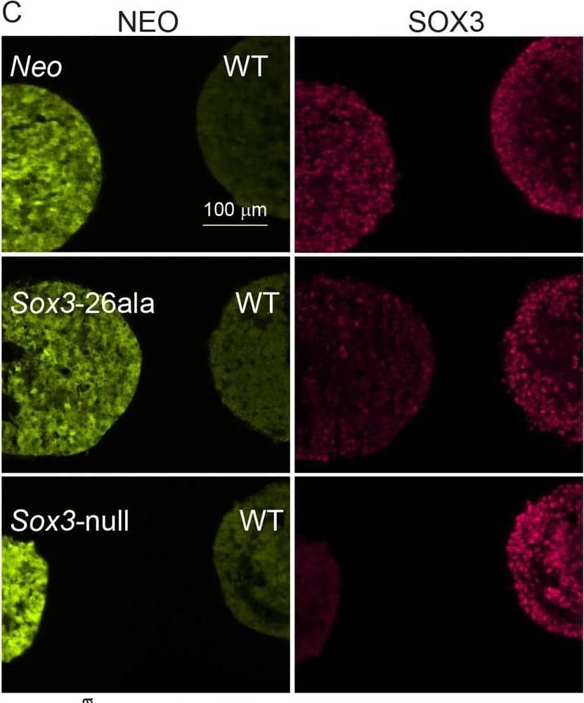
Detection of SOX3 by Immunocytochemistry/ Immunofluorescence Transcription is unaffected but protein is cleared from mutant cells.A) SOX3 protein is present in every WT cell (NEO−) of the 13.5 dpc telencephalic ventricular zone but virtually absent from equivalent tissue derived from Sox3-26ala cells (NEO+). B) Comparison of SOX3 immunostaining on Sox3-null cells (from a 14.5 dpc +/− embryo) and Sox3-26ala expressing cells (from a Sox3-26ala <-> WT chimera) confirming that the antibody is SOX3-specific and that the Sox3-26ala expressing cells exhibit a low level of residual nuclear protein. C) WT, Neo, Sox3-26ala and Sox3-null ES cells were differentiated for 5 days in CDM as multi-cellular bodies. Rare SOX3 positive cells were detected in Sox3-26ala CDMs while the majority of cells had low SOX3 protein levels in comparison to neighbouring WT CDM bodies processed on the same slide. D–E) WT, Neo, Sox3-26ala and Sox3-null ES cells were grown in N2B27 for 4 days to form neural progenitors. Western blotting for SOX3 reveals a dramatic reduction of protein in Sox3-26ala cells (D); 3 and 30 minute exposures are shown. E) Transcript levels of Sox3 are not affected in Sox3-26ala cells as determined by qPCR. Three experimental replicates are shown. Data was normalised to Sox3 levels inSox3-Neo control cells and error bars represent SEM. F) ISH confirms that Sox3 transcript is present at comparable levels in ventricular zone cells at 13.5 dpc derived from both WT (Neo−) and Sox3-26ala (Neo+) cells. ISH performed on adjacent 10 µm coronal sections of 13.5 dpc chimeric telencephalon. Image collected and cropped by CiteAb from the following open publication (https://pubmed.ncbi.nlm.nih.gov/23505376), licensed under a CC-BY license. Not internally tested by R&D Systems.
Preparation and Storage
- 12 months from date of receipt, -20 to -70 °C as supplied.
- 1 month, 2 to 8 °C under sterile conditions after reconstitution.
- 6 months, -20 to -70 °C under sterile conditions after reconstitution.
Background: SOX3
SOX3 belongs to the SOX family of transcription factors involved in the regulation of embryonic development and in the determination of cell fate. The SOX3 gene is localized on the X chromosome and is expressed in the brain and gonads. It has been suggested that SOX3 plays a role in brain formation and function. Human and mouse SOX3 share 92% amino acid sequence identity.
Product Datasheets
Citations for Human SOX3 Antibody
R&D Systems personnel manually curate a database that contains references using R&D Systems products. The data collected includes not only links to publications in PubMed, but also provides information about sample types, species, and experimental conditions.
13
Citations: Showing 1 - 10
Filter your results:
Filter by:
-
Spermatogonial fate in mice with increased activin A bioactivity and testicular somatic cell tumours
Authors: Penny A. F. Whiley, Benedict Nathaniel, Peter G. Stanton, Robin M. Hobbs, Kate L. Loveland
Frontiers in Cell and Developmental Biology
-
Chromatin remodelers HELLS, WDHD1 and BAZ1A are dynamically expressed during mouse spermatogenesis
Authors: Ram Prakash Yadav, Sini Leskinen, Lin Ma, Juho-Antti Mäkelä, Noora Kotaja
Reproduction
-
Single-cell transcriptomic landscapes of the otic neuronal lineage at multiple early embryonic ages
Authors: Y Sun, L Wang, T Zhu, B Wu, G Wang, Z Luo, C Li, W Wei, Z Liu
Cell Reports, 2022-03-22;38(12):110542.
Species: Mouse
Sample Types: Whole Cells
Applications: ICC -
The Nestin neural enhancer is essential for normal levels of endogenous Nestin in neuroprogenitors but is not required for embryo development
Authors: E Thomson, R Dawson, CH H'ng, F Adikusuma, S Piltz, PQ Thomas
PLoS ONE, 2021-11-05;16(11):e0258538.
Species: Mouse
Sample Types: Whole Tissue
Applications: IHC -
An mTORC1-dependent switch orchestrates the transition between mouse spermatogonial stem cells and clones of progenitor spermatogonia
Authors: S Suzuki, JR McCarrey, BP Hermann
Cell Reports, 2021-02-16;34(7):108752.
Species: Mouse
Sample Types: Whole Cells
Applications: Flow Cytometry, ICC -
SOX3 promotes generation of committed spermatogonia in postnatal mouse testes
Authors: D McAninch, JA Mäkelä, HM La, JN Hughes, R Lovell-Bad, RM Hobbs, PQ Thomas
Sci Rep, 2020-04-21;10(1):6751.
Species: Mouse
Sample Types: Tissue Homogenate
Applications: Immunoprecipitation -
DDX5 plays essential transcriptional and post-transcriptional roles in the maintenance and function of spermatogonia
Authors: JMD Legrand, AL Chan, HM La, FJ Rossello, ML Änkö, FV Fuller-Pac, RM Hobbs
Nat Commun, 2019;10(1):2278.
Species: Human
Sample Types: Whole Tissue
Applications: IHC -
Identification of dynamic undifferentiated cell states within the male germline
Authors: HM La, JA Mäkelä, AL Chan, FJ Rossello, CM Nefzger, JMD Legrand, M De Seram, JM Polo, RM Hobbs
Nat Commun, 2018-07-19;9(1):2819.
Species: Mouse
Sample Types: Whole Tissue
Applications: IHC -
GILZ-dependent modulation of mTORC1 regulates spermatogonial maintenance
Authors: Hue M. La, Ai-Leen Chan, Julien M. D. Legrand, Fernando J. Rossello, Christina G. Gangemi, Antonella Papa et al.
Development
-
An Eya1-Notch axis specifies bipotential epibranchial differentiation in mammalian craniofacial morphogenesis
Authors: Haoran Zhang, Li Wang, Elaine Yee Man Wong, Sze Lan Tsang, Pin-Xian Xu, Urban Lendahl et al.
eLife
Species: Mouse, Transgenic Mouse
Sample Types: Embryo
Applications: Immunohistochemistry -
Identification of Highly Conserved Putative Developmental Enhancers Bound by SOX3 in Neural Progenitors Using ChIP-Seq
Authors: Dale McAninch, Paul Thomas
PLoS ONE
-
Congenital Hydrocephalus and Abnormal Subcommissural Organ Development in Sox3 Transgenic Mice
Authors: Kristie Lee, Jacqueline Tan, Michael B. Morris, Karine Rizzoti, James Hughes, Pike See Cheah et al.
PLoS ONE
-
Identification of SOX3 as an XX male sex reversal gene in mice and humans.
Authors: Sutton E, Hughes J, White S
J. Clin. Invest., 2010-12-22;121(1):328-41.
Species: Mouse
Sample Types: Whole Tissue
Applications: IHC-Fr
FAQs
No product specific FAQs exist for this product, however you may
View all Antibody FAQsIsotype Controls
Reconstitution Buffers
Secondary Antibodies
Reviews for Human SOX3 Antibody
There are currently no reviews for this product. Be the first to review Human SOX3 Antibody and earn rewards!
Have you used Human SOX3 Antibody?
Submit a review and receive an Amazon gift card.
$25/€18/£15/$25CAN/¥75 Yuan/¥2500 Yen for a review with an image
$10/€7/£6/$10 CAD/¥70 Yuan/¥1110 Yen for a review without an image
