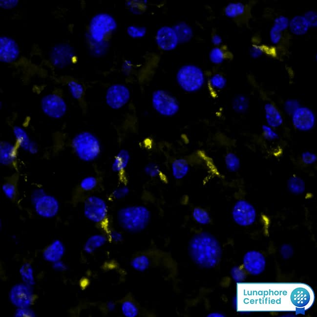
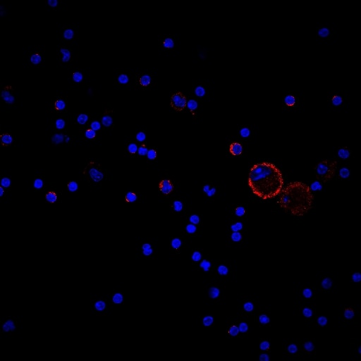
Mouse B7-1/CD80 Antibody Summary
Asp37-Lys245
Accession # Q00609
Customers also Viewed
Applications
Mouse B7-1/CD80 Sandwich Immunoassay
Please Note: Optimal dilutions should be determined by each laboratory for each application. General Protocols are available in the Technical Information section on our website.
Scientific Data
 View Larger
Detection of Mouse B7‑1/CD80 by Western Blot. Western blot shows lysates of C2C12 mouse myoblast cell line. PVDF membrane was probed with 1 µg/mL of Goat Anti-Mouse B7-1/CD80 Antigen Affinity-purified Polyclonal Antibody (Catalog # AF740) followed by HRP-conjugated Anti-Goat IgG Secondary Antibody (Catalog # HAF017). A specific band was detected for B7-1/CD80 at approximately 60 kDa (as indicated). This experiment was conducted under reducing conditions and using Immunoblot Buffer Group 1.
 View Larger
Detection of B7‑1/CD80 in Mouse Splenocytes by Flow Cytometry. Mouse splenocytes either treated with 200 ng/mL LPS (filled histogram) or unstimulated (open histogram) were stained with Goat Anti-Mouse B7-1/CD80 Antigen Affinity-purified Polyclonal Antibody (Catalog # AF740), followed by Phycoerythrin-conjugated Anti-Goat IgG Secondary Antibody (Catalog # F0107). View our protocol for Staining Membrane-associated Proteins.
 View Larger
Detection of B7‑1/CD80 in Mouse Splenocytes by Flow Cytometry Mouse B6 splenocytes treated with 200 ng/mL LPS for 48 hr were stained with (A) Goat Anti-Mouse B7-1/CD80 Antigen Affinity-purified Polyclonal Antibody (Catalog # AF740) or (B) Goat IgG control antibody (AB-108-C) followed by Phycoerythrin-conjugated Anti-Goat IgG Secondary Antibody (F0107) and Rat Anti-Mouse B220/CD45R Alexa Fluor® 488-conjugated Monoclonal Antibody (FAB1217G). View our protocol for Staining Membrane-associated Proteins.
 View Larger
IL‑2 secretion Induced by B7‑1/CD80 and Neutralization by Mouse B7‑1/CD80 Antibody. Recombinant Mouse B7-1/CD80 Fc Chimera (Catalog # 740-B1) co-stimulates IL-2 secretion in the Jurkat human acute T cell leukemia cell line in the presence of PHA in a dose-dependent manner (orange line), as measured by the Human IL-2 Quantikine ELISA Kit (Catalog # D2050). IL-2 secretion elicited by Recombinant Mouse B7-1/CD80 Fc Chimera (0.1 µg/mL) and PHA (10 µg/mL) is neutralized (green line) by increasing concentrations of Goat Anti-Mouse B7-1/CD80 Antigen Affinity-purified Polyclonal Antibody (Catalog # AF740). The ND50 is typically 0.15-0.6 µg/mL.
Preparation and Storage
- 12 months from date of receipt, -20 to -70 °C as supplied.
- 1 month, 2 to 8 °C under sterile conditions after reconstitution.
- 6 months, -20 to -70 °C under sterile conditions after reconstitution.
Background: B7-1/CD80
B7-1 and B7-2, together with their receptors CD28 and CTLA-4, constitute one of the dominant costimulatory pathways that regulate T- and B-cell responses. Although both CTLA-4 and CD28 can bind to the same ligands, CTLA-4 binds to B7-1 and B7-2 with a 20‑100 fold higher affinity than CD28 and is involved in the
down‑regulation of the immune response. B7-1 is expressed on activated B cells, activated T cells, and macrophages. B7-2 is constitutively expressed on interdigitating dendritic cells, Langerhans cells, peripheral blood dendritic cells, memory B cells, and germinal center B cells. Additionally, B7-2 is expressed at low levels on monocytes and can be up-regulated through interferon gamma. B7-1 and B7-2 are both members of the immunoglobulin superfamily. Mouse B7-1 is a 306 amino acid (aa) protein containing a putative 37 aa signal peptide, a 190 aa extracellular domain, a 22 aa transmembrane domain, and a 38 aa cytoplasmic domain. Mouse B7-1 and B7-2 share 28% amino acid identity. Mouse and human B7-1 share 44% amino acid identity. However, it has been observed that both human and mouse
B7‑1 and B7‑2 can bind to either human or mouse CD28 and CTLA-4, suggesting that there are conserved amino acids which form the B7-1/B7-2/CD28/CTLA-4 critical binding sites.
- Azuma, M. et al. (1993) Nature 366:76.
- Freeman, G.J. et al. (1993) Science 262:909.
- Freeman, G. et al. (1991) J. Exp. Med. 174:625.
- Selvakumar, A. et al. (1993) Immunogenetics 38:292.
- Chen, C. et al. (1994) J. Immunol. 152:4929.
- Freeman, G.J. et al. (1993) J. Exp. Med. 178:2185.
Product Datasheets
Citations for Mouse B7-1/CD80 Antibody
R&D Systems personnel manually curate a database that contains references using R&D Systems products. The data collected includes not only links to publications in PubMed, but also provides information about sample types, species, and experimental conditions.
11
Citations: Showing 1 - 10
Filter your results:
Filter by:
-
Systemic chromosome instability in Shugoshin-1 mice resulted in compromised glutathione pathway, activation of Wnt signaling and defects in immune system in the lung.
Authors: Yamada HY, Kumar G, Zhang Y et al.
Oncogenesis.
-
Guidelines for visualization and analysis of DC in tissues using multiparameter fluorescence microscopy imaging methods
Authors: F Bayerl, DA Bejarano, G Bertacchi, AC Doffin, E Gobbini, M Hubert, L Li, P Meiser, AM Pedde, W Posch, L Rupp, A Schlitzer, M Schmitz, BU Schraml, S Uderhardt, J Valladeau-, D Wilflingse, V Zaderer, JP Böttcher
Oncogene, 2023-01-09;0(0):e2249923.
Species: Mouse
Sample Types: Whole Tissue
Applications: IHC -
Dysregulation of Immune Response Mediators and Pain-Related Ion Channels Is Associated with Pain-like Behavior in the GLA KO Mouse Model of Fabry Disease
Authors: M Spitzel, E Wagner, M Breyer, D Henniger, M Bayin, L Hofmann, D Mauceri, C Sommer, N Üçeyler
Cells, 2022-05-24;11(11):.
Species: Mouse
Sample Types: Whole Tissue
Applications: IHC -
Receptor‑selective interleukin‑4 mutein attenuates laser‑induced choroidal neovascularization through the regulation of macrophage polarization in mice
Authors: Limo Gao, Wenmin Jiang, Haiying Liu, Zhiheng Chen, Yanhui Lin
Experimental and Therapeutic Medicine
-
Resveratrol Ameliorates Cardiac Remodeling in a Murine Model of Heart Failure With Preserved Ejection Fraction
Authors: L Zhang, J Chen, L Yan, Q He, H Xie, M Chen
Frontiers in Pharmacology, 2021-06-10;12(0):646240.
Species: Mouse
Sample Types: Whole Tissue
Applications: IHC -
High fat diet and leptin promote tumor progression by inducing myeloid-derived suppressor cells
Authors: VK Clements, T Long, R Long, C Figley, DMC Smith, S Ostrand-Ro
J. Leukoc. Biol., 2018-01-03;0(0):.
Species: Mouse
Sample Types: Whole Cells
Applications: Flow Cytometry -
TLR-mediated albuminuria needs TNF alpha -mediated cooperativity between TLRs present in hematopoietic tissues and CD80 present on non-hematopoietic tissues in mice
Authors: Nidhi Jain, Bhavya Khullar, Neelam Oswal, Balaji Banoth, Prashant Joshi, Balachandran Ravindran et al.
Disease Models & Mechanisms
Species: Mouse
Sample Types: Whole Tissue
Applications: Immunohistochemistry -
B7–1 Is Not Induced in Podocytes of Human and Experimental Diabetic Nephropathy
Authors: Elena Gagliardini, Rubina Novelli, Daniela Corna, Carlamaria Zoja, Barbara Ruggiero, Ariela Benigni et al.
Journal of the American Society of Nephrology
-
Aging is associated with increased regulatory T-cell function.
Authors: Garg S, Delaney C, Toubai T, Ghosh A, Reddy P, Banerjee R, Yung R
Aging Cell, 2014-02-25;13(3):441-8.
Species: Mouse
Sample Types: Cell Lysates
Applications: Western Blot -
Antigen-loaded ER microsomes from APC induce potent immune responses against viral infection.
Authors: Sofra V, Mansour S, Liu M, Gao B, Primpidou E, Wang P, Li S
Eur. J. Immunol., 2009-01-01;39(1):85-95.
Species: Mouse
Sample Types: Tissue Homogenates
Applications: Western Blot -
Suppression of activation and induction of apoptosis in RAW264.7 cells by amniotic membrane extract.
Authors: He H, Li W, Chen SY, Zhang S, Chen YT, Hayashida Y, Zhu YT, Tseng SC
Invest. Ophthalmol. Vis. Sci., 2008-07-09;49(10):4468-75.
Species: Mouse
Sample Types: Whole Cells
Applications: ICC
FAQs
No product specific FAQs exist for this product, however you may
View all Antibody FAQsIsotype Controls
Reconstitution Buffers
Secondary Antibodies
Reviews for Mouse B7-1/CD80 Antibody
Average Rating: 5 (Based on 2 Reviews)
Have you used Mouse B7-1/CD80 Antibody?
Submit a review and receive an Amazon gift card.
$25/€18/£15/$25CAN/¥75 Yuan/¥2500 Yen for a review with an image
$10/€7/£6/$10 CAD/¥70 Yuan/¥1110 Yen for a review without an image
Filter by:


