Mouse CDO Antibody Summary
Val22-Leu963
Accession # AAC43031
Customers also Viewed
Applications
Please Note: Optimal dilutions should be determined by each laboratory for each application. General Protocols are available in the Technical Information section on our website.
Scientific Data
 View Larger
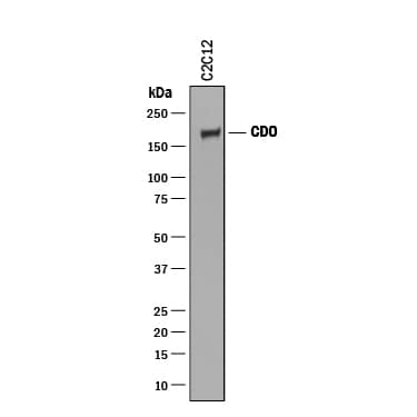
Detection of Mouse CDO by Western Blot. Western blot shows lysate of C2C12 mouse myoblast cell line. PVDF membrane was probed with 0.25 µg/mL of Goat Anti-Mouse CDO Antigen Affinity-purified Polyclonal Antibody (Catalog # AF2429) followed by HRP-conjugated Anti-Goat IgG Secondary Antibody (Catalog # HAF019). A specific band was detected for CDO at approximately 190 kDa (as indicated). This experiment was conducted under reducing conditions and using Immunoblot Buffer Group 1.
 View Larger
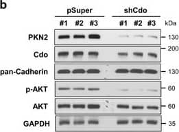
Detection of Mouse CDO by Knockdown Validated PKN2 levels are elevated during myoblast differentiation and decreased in Cdo-depleted cells with a concurrent reduction in AKT activation. (a) C2C12 cells were cultured to near confluency (D0) and induced to differentiate in differentiation medium (DM) for total 3 days (D3). Lysates were immunoblotted with antibodies to PKN2, Cdo, MHC, MyoD, myogenin, phosphorylated-AKT (p-AKT) and AKT. GAPDH and pan-Cadherin serve as loading controls. (b) Lysates of C2C12 cells transiently transfected with Cdo or control expression vectors as indicated were immunoblotted with antibodies to PKN2, Cdo, p-AKT and AKT. GAPDH and pan-Cadherin serve as loading controls. (c) Immunoblot analysis for the expression of PKN2, p-AKT and AKT proteins in Cdo+/+ and Cdo−/− primary myoblasts from hindlimb muscles, and pan-Cadherin serves as a loading control. (d) Photomicrographs of C2C12 cells that stably express PKN2 or control vectors were cultured in DM for 2 days, fixed, and immunostained with an antibody to MHC followed by DAPI staining to visualize nuclei. Size bar, 100 μm. (e) Quantification of myotube formation shown in (d). Values represent means of triplicate determinations ±1 S.D. The experiment was repeated three times with similar results. Significant difference from control, *P<0.01. (f) C2C12 cells stably transfected with shPKN2 or control (pSuper) vectors, and cultured to confluency and induced to differentiate for total 3 days. Cell lysates were immunoblotted using antibodies to PKN2, MHC, MyoD, Myogenin and pan-Cadherin as a loading control. (g) Photomicrographs of C2C12 cells stably transfected with PKN2 shRNA or control vectors were cultured in DM for 3 days, fixed and immunostained with an antibody to MHC followed by DAPI staining to visualize nuclei. Size bar, 100 μm. (h) Quantification of myotube formation by cell lines shown in (g). Values represent means of triplicate determinations ±1 S.D. The experiment was repeated three times with similar results. Significant difference from control, *P<0.01 Image collected and cropped by CiteAb from the following publication (https://www.nature.com/articles/cddis2016296), licensed under a CC-BY license. Not internally tested by R&D Systems.
 View Larger
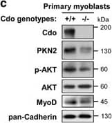
Detection of Mouse CDO by Knockout Validated PKN2 levels are elevated during myoblast differentiation and decreased in Cdo-depleted cells with a concurrent reduction in AKT activation. (a) C2C12 cells were cultured to near confluency (D0) and induced to differentiate in differentiation medium (DM) for total 3 days (D3). Lysates were immunoblotted with antibodies to PKN2, Cdo, MHC, MyoD, myogenin, phosphorylated-AKT (p-AKT) and AKT. GAPDH and pan-Cadherin serve as loading controls. (b) Lysates of C2C12 cells transiently transfected with Cdo or control expression vectors as indicated were immunoblotted with antibodies to PKN2, Cdo, p-AKT and AKT. GAPDH and pan-Cadherin serve as loading controls. (c) Immunoblot analysis for the expression of PKN2, p-AKT and AKT proteins in Cdo+/+ and Cdo−/− primary myoblasts from hindlimb muscles, and pan-Cadherin serves as a loading control. (d) Photomicrographs of C2C12 cells that stably express PKN2 or control vectors were cultured in DM for 2 days, fixed, and immunostained with an antibody to MHC followed by DAPI staining to visualize nuclei. Size bar, 100 μm. (e) Quantification of myotube formation shown in (d). Values represent means of triplicate determinations ±1 S.D. The experiment was repeated three times with similar results. Significant difference from control, *P<0.01. (f) C2C12 cells stably transfected with shPKN2 or control (pSuper) vectors, and cultured to confluency and induced to differentiate for total 3 days. Cell lysates were immunoblotted using antibodies to PKN2, MHC, MyoD, Myogenin and pan-Cadherin as a loading control. (g) Photomicrographs of C2C12 cells stably transfected with PKN2 shRNA or control vectors were cultured in DM for 3 days, fixed and immunostained with an antibody to MHC followed by DAPI staining to visualize nuclei. Size bar, 100 μm. (h) Quantification of myotube formation by cell lines shown in (g). Values represent means of triplicate determinations ±1 S.D. The experiment was repeated three times with similar results. Significant difference from control, *P<0.01 Image collected and cropped by CiteAb from the following publication (https://www.nature.com/articles/cddis2016296), licensed under a CC-BY license. Not internally tested by R&D Systems.
Preparation and Storage
- 12 months from date of receipt, -20 to -70 °C as supplied.
- 1 month, 2 to 8 °C under sterile conditions after reconstitution.
- 6 months, -20 to -70 °C under sterile conditions after reconstitution.
Background: CDO
Mouse CDO is a 190 kDa member of the Ig/FN-type III repeat subfamily of transmembrane (TM) proteins. The extracellular region contains five Ig-like domains and three FN-III modules. The molecule forms cis-complexes with itself and the related molecule termed BOC. Mouse CDO extracellular domain shares 95% and 85% aa identity with the corresponding regions of rat and human CDO, respectively.
Product Datasheets
Citations for Mouse CDO Antibody
R&D Systems personnel manually curate a database that contains references using R&D Systems products. The data collected includes not only links to publications in PubMed, but also provides information about sample types, species, and experimental conditions.
12
Citations: Showing 1 - 10
Filter your results:
Filter by:
-
Differential Expression of NF2 in Neuroepithelial Compartments Is Necessary for Mammalian Eye Development.
Authors: Moon K H, Kim H T et al.
Dev Cell
-
MiR-193b-365, a brown fat enriched microRNA cluster, is essential for brown fat differentiation
Authors: Lei Sun, Huangming Xie, Marcelo A. Mori, Ryan Alexander, Bingbing Yuan, Shilpa M. Hattangadi et al.
Nature Cell Biology
-
Mesocortical Dopamine Phenotypes in Mice Lacking the Sonic Hedgehog Receptor Cdon
Authors: Michael Verwey, Alanna Grant, Nicholas Meti, Lauren Adye-White, Angelica Torres-Berrío, Veronique Rioux et al.
eNeuro
-
Distinct structural requirements for CDON and BOC in the promotion of Hedgehog signaling
Authors: Jane Y. Song, Alexander M. Holtz, Justine M. Pinskey, Benjamin L. Allen
Developmental Biology
-
Lhx2 is a progenitor-intrinsic modulator of Sonic Hedgehog signaling during early retinal neurogenesis
Authors: X Li, PJ Gordon, JA Gaynes, AW Fuller, R Ringuette, CP Santiago, V Wallace, S Blackshaw, P Li, EM Levine
Elife, 2022-12-02;11(0):.
Species: Mouse
Sample Types: Whole Tissue
Applications: IHC -
mTORC1-induced retinal progenitor cell overproliferation leads to accelerated mitotic aging and degeneration of descendent M�ller glia
Authors: S Lim, YJ Kim, S Park, JH Choi, Y Sung, K Nishimori, Z Kozmik, HW Lee, JW Kim
Elife, 2021-10-22;10(0):.
Species: Mouse
Sample Types: Whole Tissue
Applications: IHC -
ZSWIM8 is a myogenic protein that partly prevents C2C12 differentiation
Authors: F Okumura, N Oki, Y Fujiki, R Ikuta, K Osaki, S Hamada, K Nakatsukas, N Hisamoto, T Hara, T Kamura
Scientific Reports, 2021-10-22;11(1):20880.
Species: Mouse
Sample Types: Cell Lysates, Whole Cells
Applications: ICC, Western Blot -
Dehydrocorydaline promotes myogenic differentiation via p38�MAPK activation
Mol Med Rep, 2016-08-19;14(4):3029-36.
Species: Mouse
Sample Types: Cell Lysates
Applications: Western Blot -
Syntaxin 4 regulates the surface localization of a promyogenic receptor Cdo thereby promoting myogenic differentiation.
Authors: Yoo M, Kim B, Lee S, Jeong H, Park J, Seo D, Kim Y, Lee H, Han J, Kang J, Bae G
Skelet Muscle, 2015-09-07;5(0):28.
Species: Mouse
Sample Types: Cell Lysates
Applications: Western Blot -
Segregation of ipsilateral retinal ganglion cell axons at the optic chiasm requires the Shh receptor Boc.
Authors: Fabre PJ, Shimogori T, Charron F
J. Neurosci., 2010-01-06;30(0):266.
Species: Mouse
Sample Types: Whole Tissue
Applications: IHC-P -
Desert Hedgehog-Driven Endothelium Integrity Is Enhanced by Gas1 (Growth Arrest-Specific 1) but Negatively Regulated by Cdon (Cell Adhesion Molecule-Related/Downregulated by Oncogenes)
Authors: Candice Chapouly, Pierre-Louis Hollier, Sarah Guimbal, Lauriane Cornuault, Alain-Pierre Gadeau, Marie-Ange Renault
Arteriosclerosis, Thrombosis, and Vascular Biology
-
PKN2 and Cdo interact to activate AKT and promote myoblast differentiation
Authors: Sang-Jin Lee, Jeongmi Hwang, Hyeon-Ju Jeong, Miran Yoo, Ga-Yeon Go, Jae-Rin Lee et al.
Cell Death & Disease
FAQs
No product specific FAQs exist for this product, however you may
View all Antibody FAQsIsotype Controls
Reconstitution Buffers
Secondary Antibodies
Reviews for Mouse CDO Antibody
There are currently no reviews for this product. Be the first to review Mouse CDO Antibody and earn rewards!
Have you used Mouse CDO Antibody?
Submit a review and receive an Amazon gift card.
$25/€18/£15/$25CAN/¥75 Yuan/¥2500 Yen for a review with an image
$10/€7/£6/$10 CAD/¥70 Yuan/¥1110 Yen for a review without an image


