Mouse/Rat FABP2/I-FABP Antibody Summary
Met1-Glu132
Accession # P02693
Customers also Viewed
Applications
Please Note: Optimal dilutions should be determined by each laboratory for each application. General Protocols are available in the Technical Information section on our website.
Scientific Data
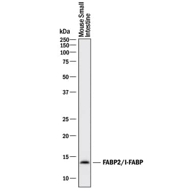 View Larger
Detection of Mouse FABP2/I‑FABP by Western Blot. Western blot shows lysates of mouse small intestine tissue. PVDF membrane was probed with 1 µg/mL of Goat Anti-Mouse/Rat FABP2/I-FABP Antigen Affinity-purified Polyclonal Antibody (Catalog # AF1486) followed by HRP-conjugated Anti-Goat IgG Secondary Antibody (Catalog # HAF017). A specific band was detected for FABP2/I-FABP at approximately 14 kDa (as indicated). This experiment was conducted under reducing conditions and using Immunoblot Buffer Group 1.
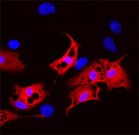 View Larger
FABP2/I‑FABP in Rat Mesenchymal Stem Cells. FABP2/I-FABP was detected in immersion fixed rat mesenchymal stem cells differentiated to adipocytes using Goat Anti-Mouse/Rat FABP2/I-FABP Antigen Affinity-purified Polyclonal Antibody (Catalog # AF1486) at 10 µg/mL for 3 hours at room temperature. Cells were stained using the NorthernLights™ 557-conjugated Anti-Goat IgG Secondary Antibody (red; Catalog # NL001) and counterstained with DAPI (blue). Specific staining was localized to cytoplasm. View our protocol for Fluorescent ICC Staining of Stem Cells on Coverslips.
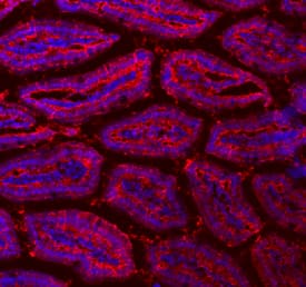 View Larger
FABP2/I‑FABP in Mouse Intestine. FABP2/I-FABP was detected in immersion fixed frozen sections of adult mouse intestine using Goat Anti-Mouse/Rat FABP2/I-FABP Antigen Affinity-purified Polyclonal Antibody (Catalog # AF1486) at 10 µg/mL overnight at 4 °C. Tissue was stained using the NorthernLights™ 557-conjugated Anti-Goat IgG Secondary Antibody (red; Catalog # NL001) and counterstained with DAPI (blue). Specific staining was localized to intestinal epithelia. View our protocol for Fluorescent IHC Staining of Frozen Tissue Sections.
 View Larger
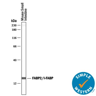
Detection of Mouse FABP2/I‑FABP by Simple WesternTM. Simple Western lane view shows lysates of mouse small intestine tissue, loaded at 0.2 mg/mL. A specific band was detected for FABP2/I-FABP at approximately 18 kDa (as indicated) using 20 µg/mL of Goat Anti-Mouse/Rat FABP2/I-FABP Antigen Affinity-purified Polyclonal Antibody (Catalog # AF1486) followed by 1:50 dilution of HRP-conjugated Anti-Goat IgG Secondary Antibody (Catalog # HAF019). This experiment was conducted under reducing conditions and using the 12-230 kDa separation system.
Preparation and Storage
- 12 months from date of receipt, -20 to -70 °C as supplied.
- 1 month, 2 to 8 °C under sterile conditions after reconstitution.
- 6 months, -20 to -70 °C under sterile conditions after reconstitution.
Background: FABP2/I-FABP
FABP2, also named I-FABP and gFABP, is a member of the intracellular fatty acid binding protein family. It is highly expressed in the intestine. FABP2 binds fatty acid in a non-covalent 1:1 complex to chaperone the lipids to cellular enzymes for metabolism and signal transduction.
Product Datasheets
Citation for Mouse/Rat FABP2/I-FABP Antibody
R&D Systems personnel manually curate a database that contains references using R&D Systems products. The data collected includes not only links to publications in PubMed, but also provides information about sample types, species, and experimental conditions.
1 Citation: Showing 1 - 1
-
Nkx2.2 is expressed in a subset of enteroendocrine cells with expanded lineage potential.
Authors: Gross S, Balderes D, Liu J et al.
Am J Physiol Gastrointest Liver Physiol
FAQs
No product specific FAQs exist for this product, however you may
View all Antibody FAQsIsotype Controls
Reconstitution Buffers
Secondary Antibodies
Reviews for Mouse/Rat FABP2/I-FABP Antibody
There are currently no reviews for this product. Be the first to review Mouse/Rat FABP2/I-FABP Antibody and earn rewards!
Have you used Mouse/Rat FABP2/I-FABP Antibody?
Submit a review and receive an Amazon gift card.
$25/€18/£15/$25CAN/¥75 Yuan/¥2500 Yen for a review with an image
$10/€7/£6/$10 CAD/¥70 Yuan/¥1110 Yen for a review without an image


