Rat MuSK Antibody Summary
Customers also Viewed
Applications
Please Note: Optimal dilutions should be determined by each laboratory for each application. General Protocols are available in the Technical Information section on our website.
Scientific Data
 View Larger
Acetylcholine Receptor Clustering Mediated by MuSK and Neutralization by Rat MuSK Antibody. Recombinant Rat Agrin (Catalog # 550-AG) induces MuSK-dependent acetylcholine receptor clustering on myotubes differentiated from the C2C12 mouse myoblast cell line in a dose-dependent manner (orange line). Acetylcholine Receptor Clustering elicited by Recombinant Rat Agrin (16 ng/mL) is neutralized (green line) by increasing concentrations of Goat Anti-Rat MuSK Antigen Affinity-purified Polyclonal Antibody (Catalog # AF562). The ND50 is typically 0.5-2 µg/mL.
 View Larger
Detection of Human MuSK by Immunocytochemistry/ Immunofluorescence MuSK patient IgG4 or IgG1-3 do not induce endocytosis of MuSK.Patient plasma or purified IgG1-3 or IgG4 were applied at 0.17nM final concentration of MuSK antibody to HEK293 cells expressing MuSK at the cell surface. Cells were either incubated at 4°C to prevent, or at 37°C to allow, endocytosis. Patient antibody binding was visualised by addition of a secondary fluorescent anti-human antibody at the end of the experiment. (A) An example of cells that were incubated with IgG1-3 from patient 9. Robust staining was observed after 6 hours incubation at both temperatures. Scale bar=25µm. (B) Staining was scored by two individuals as described in methods, and the scores were normalised to the score at 0 hours for each condition. A slight decrease in staining was observed for cells incubated at 37°C, and pre-fixed cells also showed this reduction. (C) As a positive control for endocytosis, IgG1-3 or IgG4 was cross-linked by addition of Alexa Fluor 568-conjugated anti-human IgG prior to incubation. Example images show cells treated with patient 9 IgG4 and IgG1-3 in the presence and absence of cross-linking anti-human IgG after 6 hours incubation at 37°C. There is a clear difference when the cross-linking secondary antibody is present. Scale bar =50µm (D) Cross-linking by the secondary antibody induced internalisation of human anti-MuSK antibodies as well as MuSK-EGFP as early as 30 minutes, as observed by confocal microscopy. An example image of a cell from a confocal z-stack with orthogonal side-views is shown. The arrow shows internalised secondary Alexa Fluor 568-conjugated anti-human IgG (red) colocalised with EGFP-tagged MuSK (green). Scale bar =5µm. Image collected and cropped by CiteAb from the following open publication (https://dx.plos.org/10.1371/journal.pone.0080695), licensed under a CC-BY license. Not internally tested by R&D Systems.
 View Larger
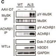
Detection of Mouse MuSK by Western Blot DOK7 gene therapy suppresses motor nerve terminal degeneration at the NMJ in ALS miceAForelimb grip strength of wild‐type (WT) mice&that of control&AAV‐D7 treatment groups of ALS mice at P84 (WT, n = 5 mice; control group, n = 5 mice; treatment group, n = 6 mice). Values are means ± SEM. **P = 0.0016 (WT vs. control group)&0.0003 (WT vs. treatment group) by one‐way analysis of variance (ANOVA) with Bonferroni's post hoc test. n.s., not significant.BThe difference in cycle threshold ( delta Ct) between the human SOD1‐G93A transgene&the reference mouse apob gene. To calculate the human transgene level, the delta Ct value of hSOD1 was subtracted from the delta Ct value of apob (control group, n = 5 mice; treatment group, n = 6 mice).C–GWT or ALS mice treated or not with 1.2 × 10¹² vg of AAV‐D7 at P90 subjected to the following assays at P120. Tyrosine phosphorylation of MuSK or AChR in the hind‐limb muscle. MuSK or AChR beta 1 subunit (AChR beta 1) immunoprecipitates (IP) from whole‐tissue lysates (WTLs) of the hind‐limb muscle immunoblotted for phosphotyrosine (pY), MuSK,&AChR beta 1. WTLs blotted for Dok‐7‐EGFP, human SOD1,&GAPDH (C). Whole‐mount staining of NMJs on the diaphragm muscle. Motor nerve terminals (green)&postsynaptic AChRs (red) stained with anti‐synapsin‐1 antibodies& alpha ‐bungarotoxin, respectively. Expression of Dok‐7‐EGFP fusion protein (gray) was monitored by EGFP. Scale bars, 50 μm. NT, not treated (D). The size of AChR cluster (E), the size of motor nerve terminal (F),&innervation ratio (G) (WT‐NT, n = 5 mice; ALS‐NT, n = 5 mice; ALS‐AAV‐D7, n = 6 mice). Values are means ± SD. (E) ***P < 0.0001; (F) **P = 0.0077, ***P = 0.0001; (G) *P = 0.0134, ***P < 0.0001 by one‐way ANOVA with Dunnett's post hoc test.Source data are available online for this figure. Image collected&cropped by CiteAb from the following open publication (https://pubmed.ncbi.nlm.nih.gov/28490573), licensed under a CC-BY license. Not internally tested by R&D Systems.
Preparation and Storage
- 12 months from date of receipt, -20 to -70 °C as supplied.
- 1 month, 2 to 8 °C under sterile conditions after reconstitution.
- 6 months, -20 to -70 °C under sterile conditions after reconstitution.
Background: MuSK
MuSK (Muscle-Specific Kinase) is a receptor tyrosine kinase expressed in muscle that regulates the formation of the neuromuscular junction. It binds the heparin sulfate proteoglycan Agrin to promote Acetylcholine Receptor clustering.
Product Datasheets
Citations for Rat MuSK Antibody
R&D Systems personnel manually curate a database that contains references using R&D Systems products. The data collected includes not only links to publications in PubMed, but also provides information about sample types, species, and experimental conditions.
16
Citations: Showing 1 - 10
Filter your results:
Filter by:
-
MuSK myasthenia gravis monoclonal antibodies: Valency dictates pathogenicity
Authors: Maartje G. Huijbers, Dana L. Vergoossen, Yvonne E. Fillié-Grijpma, Inge E. van Es, Marvyn T. Koning, Linda M. Slot et al.
Neurology - Neuroimmunology Neuroinflammation
-
Development and characterization of agonistic antibodies targeting the Ig-like 1 domain of MuSK
Authors: Lim, JL;Augustinus, R;Plomp, JJ;Roya-Kouchaki, K;Vergoossen, DLE;Filli�-Grijpma, Y;Struijk, J;Thomas, R;Salvatori, D;Steyaert, C;Blanchetot, C;Vanhauwaert, R;Silence, K;van der Maarel, SM;Verschuuren, JJ;Huijbers, MG;
Scientific reports
Species: Mouse
Sample Types: Cell Lysates
Applications: Western Blot -
Mechanism of disease and therapeutic rescue of Dok7 congenital myasthenia
Authors: J Oury, W Zhang, N Leloup, A Koide, AD Corrado, G Ketavarapu, T Hattori, S Koide, SJ Burden
Nature, 2021-06-23;0(0):.
Species: Mouse
Sample Types: Cell Culture Supernates
Applications: Western Blot -
Characterization of pathogenic monoclonal autoantibodies derived from muscle-specific kinase myasthenia gravis patients
Authors: K Takata, P Stathopoul, M Cao, M Mané-Damas, ML Fichtner, ES Benotti, L Jacobson, P Waters, SR Irani, P Martinez-M, D Beeson, M Losen, A Vincent, RJ Nowak, KC O'Connor
JCI Insight, 2019-06-20;4(12):.
Species: Mouse
Sample Types: Cell Lysates
Applications: Immunoprecipitation, Western Blot -
DOK7 gene therapy enhances motor activity and life span in ALS model mice
Authors: S Miyoshi, T Tezuka, S Arimura, T Tomono, T Okada, Y Yamanashi
EMBO Mol Med, 2017-07-01;0(0):.
Species: Mouse
Sample Types: Tissue Homogenates
Applications: Western Blot -
IgG-specific cell-based assay detects potentially pathogenic MuSK-Abs in seronegative MG
Authors: S Huda, P Waters, M Woodhall, MI Leite, L Jacobson, A De Rosa, M Maestri, R Ricciardi, JM Heckmann, A Maniaol, A Evoli, J Cossins, D Hilton-Jon, A Vincent
Neurol Neuroimmunol Neuroinflamm, 2017-06-05;4(4):e357.
Species: Human
Sample Types: Whole Cells
Applications: ICC -
Laminin is Instructive and Calmodulin Dependent Kinase II is Non-Permissive for the Formation of Complex Aggregates of Acetylcholine Receptors on Myotubes in Culture
Authors: Raphael Vezina-Aud
Matrix Biol, 2016-12-10;0(0):.
Species: Mouse
Sample Types: Whole Cells
Applications: Neutralization -
MuSK myasthenia gravis IgG4 disrupts the interaction of LRP4 with MuSK but both IgG4 and IgG1-3 can disperse preformed agrin-independent AChR clusters.
Authors: Koneczny, Inga, Cossins, Judith, Waters, Patrick, Beeson, David, Vincent, Angela
PLoS ONE, 2013-11-07;8(11):e80695.
Species: Human
Sample Types: Cell Lysates
Applications: Immunoprecipitation -
Immunocapture and identification of cell membrane protein antigenic targets of serum autoantibodies.
Authors: Littleton E, Dreger M, Palace J
Mol. Cell Proteomics, 2009-03-29;8(7):1688-96.
Species: Human
Sample Types: Cell Lysates
Applications: Western Blot -
Mutations causing DOK7 congenital myasthenia ablate functional motifs in Dok-7.
Authors: Hamuro J, Higuchi O, Okada K, Ueno M, Iemura S, Natsume T, Spearman H, Beeson D, Yamanashi Y
J. Biol. Chem., 2007-12-29;283(9):5518-24.
Species: Mouse
Sample Types: Cell Culture Supernates
Applications: Western Blot -
MuSK expressed in the brain mediates cholinergic responses, synaptic plasticity, and memory formation.
Authors: Garcia-Osta A, Tsokas P, Pollonini G, Landau EM, Blitzer R, Alberini CM
J. Neurosci., 2006-07-26;26(30):7919-32.
Species: Mouse, Rat
Sample Types: Cell Lysates, Tissue Homogenates
Applications: Western Blot -
14-3-3 gamma associates with muscle specific kinase and regulates synaptic gene transcription at vertebrate neuromuscular synapse.
Authors: Strochlic L, Cartaud A, Mejat A, Grailhe R, Schaeffer L, Changeux JP, Cartaud J
Proc. Natl. Acad. Sci. U.S.A., 2004-12-16;101(52):18189-94.
Species: Rat
Sample Types: Cell Lysates
Applications: Immunoprecipitation, Western Blot -
Postsynaptic requirement for Abl kinases in assembly of the neuromuscular junction.
Authors: Finn AJ, Feng G, Pendergast AM
Nat. Neurosci., 2003-07-01;6(7):717-23.
Species: Mouse
Sample Types: Cell Lysates
Applications: Immunoprecipitation, Western Blot -
The MuSK activator agrin has a separate role essential for postnatal maintenance of neuromuscular synapses
Authors: Tohru Tezuka, Akane Inoue, Taisuke Hoshi, Scott D. Weatherbee, Robert W. Burgess, Ryo Ueta et al.
Proceedings of the National Academy of Sciences
-
IgG1-3 MuSK Antibodies Inhibit AChR Cluster Formation, Restored by SHP2 Inhibitor, Despite Normal MuSK, DOK7, or AChR Subunit Phosphorylation
Authors: Michelangelo Cao, Wei-Wei Liu, Susan Maxwell, Saif Huda, Richard Webster, Amelia Evoli et al.
Neurology - Neuroimmunology Neuroinflammation
-
SHP2 inhibitor protects AChRs from effects of myasthenia gravis MuSK antibody
Authors: Saif Huda, Michelangelo Cao, Anna De Rosa, Mark Woodhall, Pedro M. Rodriguez Cruz, Judith Cossins et al.
Neurology - Neuroimmunology Neuroinflammation
FAQs
No product specific FAQs exist for this product, however you may
View all Antibody FAQsIsotype Controls
Reconstitution Buffers
Secondary Antibodies
Reviews for Rat MuSK Antibody
There are currently no reviews for this product. Be the first to review Rat MuSK Antibody and earn rewards!
Have you used Rat MuSK Antibody?
Submit a review and receive an Amazon gift card.
$25/€18/£15/$25CAN/¥75 Yuan/¥2500 Yen for a review with an image
$10/€7/£6/$10 CAD/¥70 Yuan/¥1110 Yen for a review without an image


