Human FoxP3 Antibody Summary
Gln105-Lys200
Accession # Q9BZS1
Customers also Viewed
Applications
Please Note: Optimal dilutions should be determined by each laboratory for each application. General Protocols are available in the Technical Information section on our website.
Scientific Data
 View Larger
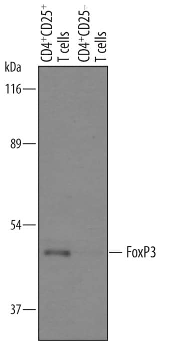
Detection of Human FoxP3 by Western Blot. Western blot shows lysates of human CD4+CD25+T cells and human CD4+CD25-T cells. PVDF membrane was probed with 1 µg/mL of Goat Anti-Human FoxP3 Antigen Affinity-purified Polyclonal Antibody (Catalog # AF3240) followed by HRP-conjugated Anti-Goat IgG Secondary Antibody (Catalog # HAF019). A specific band was detected for FoxP3 at approximately 47 kDa (as indicated). This experiment was conducted under reducing conditions and using Immunoblot Buffer Group 8.
 View Larger
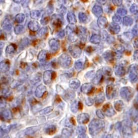
FoxP3 in Human Tonsil. FoxP3 was detected in immersion fixed paraffin-embedded sections of human tonsil using 10 µg/mL Goat Anti-Human FoxP3 Antigen Affinity-purified Polyclonal Antibody (Catalog # AF3240) overnight at 4 °C. Tissue was stained with the Anti-Goat HRP-DAB Cell & Tissue Staining Kit (brown; Catalog # CTS008) and counterstained with hematoxylin (blue). View our protocol for Chromogenic IHC Staining of Paraffin-embedded Tissue Sections.
 View Larger
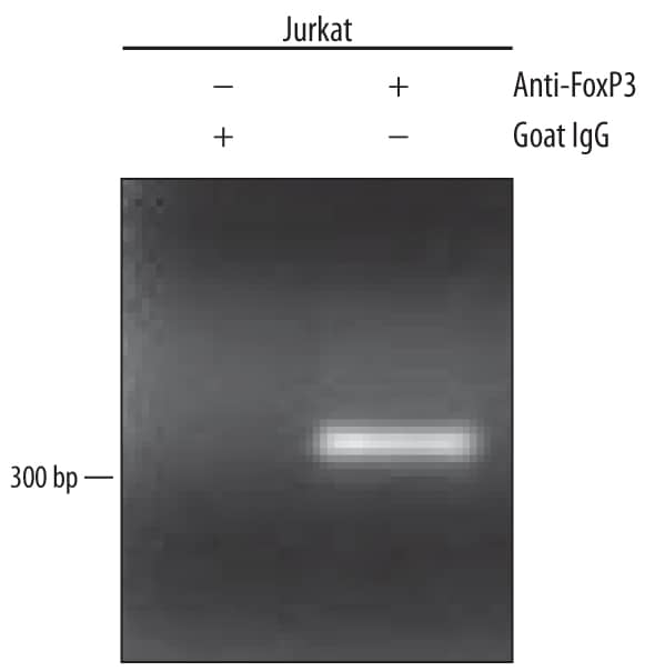
Detection of FoxP3-regulated Genes by Chromatin Immunoprecipitation. Jurkat human acute T cell leukemia cell line treated with 50 ng/mL PMA and 200 ng/mL calcium ionomycin overnight was fixed using formaldehyde, resuspended in lysis buffer, and sonicated to shear chromatin. FoxP3/DNA complexes were immunoprecipitated using 5 µg Goat Anti-Human FoxP3 Antigen Affinity-purified Polyclonal Antibody (Catalog # AF3240) or control antibody (Catalog # AB-108-C) for 15 minutes in an ultrasonic bath, followed by Biotinylated Anti-Goat IgG Secondary Antibody (Catalog # BAF109). Immunocomplexes were captured using 50 µL of MagCellect Streptavidin Ferrofluid (Catalog # MAG999) and DNA was purified using chelating resin solution. TheIL-2promoter was detected by standard PCR.
Preparation and Storage
- 12 months from date of receipt, -20 to -70 °C as supplied.
- 1 month, 2 to 8 °C under sterile conditions after reconstitution.
- 6 months, -20 to -70 °C under sterile conditions after reconstitution.
Background: FoxP3
Human FoxP3 is a 47 kDa member of the P subclass of the FOX (forkhead box) family of transcription factors. It contains a Leu-rich repeat, a C2H2 zinc finger region, and a C-terminal FKH (fork head), DNA-binding domain. Three isoforms for FoxP3 have been reported. All three isoforms share the sequence used as the immunogen. FoxP3 directly associates with NFAT and NFkB, suppressing their activity in CD4+ T cells. In human, FoxP3 is found in CD4+, CD8+ and CD4+CD25+ T cells. Over the region used for immunization of the amino acid sequence, mouse FoxP3 is 83% to 88% identical to rat, human, canine, and bovine FoxP3.
Product Datasheets
Citations for Human FoxP3 Antibody
R&D Systems personnel manually curate a database that contains references using R&D Systems products. The data collected includes not only links to publications in PubMed, but also provides information about sample types, species, and experimental conditions.
4
Citations: Showing 1 - 4
Filter your results:
Filter by:
-
Nanoscale imaging of clinical specimens using conventional and rapid-expansion pathology
Authors: Octavian Bucur, Feifei Fu, Mike Calderon, Geetha H. Mylvaganam, Ngoc L. Ly, Jimmy Day et al.
Nature Protocols
-
Tumor Lymphocyte Infiltration Is Correlated with a Favorable Tumor Regression Grade after Neoadjuvant Treatment for Esophageal Adenocarcinoma
Authors: R Haddad, O Zlotnik, T Goshen-Lag, M Levi, E Brook, B Brenner, Y Kundel, I Ben-Aharon, H Kashtan
Journal of personalized medicine, 2022-04-13;12(4):.
Species: Human
Sample Types: Whole Tissue
Applications: IHC -
Nuclear galectin-1-FOXP3 interaction dampens the tumor-suppressive properties of FOXP3 in breast cancer
Authors: Y Gao, X Li, Z Shu, K Zhang, X Xue, W Li, Q Hao, Z Wang, W Zhang, S Wang, C Zeng, D Fan, W Zhang, Y Zhang, H Zhao, M Li, C Zhang
Cell Death Dis, 2018-04-01;9(4):416.
Species: Human
Sample Types: Whole Cells
Applications: Flow Cytometry -
Human CD4+ HLA-G+ regulatory T cells are potent suppressors of graft-versus-host disease in vivo.
Authors: Pankratz S, Bittner S, Herrmann A, Schuhmann M, Ruck T, Meuth S, Wiendl H
FASEB J, 2014-04-17;28(8):3435-45.
Species: Human
Sample Types: Cell Lysates
Applications: Western Blot
FAQs
No product specific FAQs exist for this product, however you may
View all Antibody FAQsIsotype Controls
Reconstitution Buffers
Secondary Antibodies
Reviews for Human FoxP3 Antibody
There are currently no reviews for this product. Be the first to review Human FoxP3 Antibody and earn rewards!
Have you used Human FoxP3 Antibody?
Submit a review and receive an Amazon gift card.
$25/€18/£15/$25CAN/¥75 Yuan/¥2500 Yen for a review with an image
$10/€7/£6/$10 CAD/¥70 Yuan/¥1110 Yen for a review without an image


