Human BAFF/BLyS/TNFSF13B Antibody Summary
Ala134-Leu285
Accession # Q9Y275
Customers also Viewed
Applications
Please Note: Optimal dilutions should be determined by each laboratory for each application. General Protocols are available in the Technical Information section on our website.
Scientific Data
 View Larger
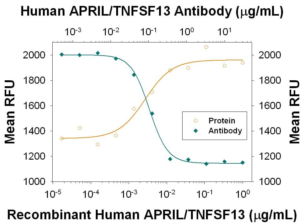
Cell Proliferation Induced By BAFF/BLyS/TNFSF13B and Neutralization by Human BAFF/BLyS/TNFSF13B Antibody. In the presence of Goat F(ab')2 Anti-mouse IgM (1 µg/mL), Recombinant Human BAFF/BLyS/TNFSF13B (Catalog # 2149-BF) stimulates proliferation in mouse B cells in a dose-dependent manner (orange line). Under these conditions, proliferation elicited by Recombinant Human BAFF/BLyS/TNFSF13B (5 ng/mL) is neutralized (green line) by increasing concentrations of Goat Anti-Human BAFF/BLyS/TNFSF13B Antigen Affinity-purified Polyclonal Antibody (Catalog # AF124). The ND50 is typically 3-12 ng/mL.
 View Larger
BAFF/BLyS/TNFSF13B in Human Spleen. BAFF/BLyS/TNFSF13B was detected in formalin fixed paraffin-embedded sections of human spleen using 15 µg/mL Goat Anti-Human BAFF/BLyS/TNFSF13B Antigen Affinity-purified Polyclonal Antibody (Catalog # AF124) overnight at 4 °C. Tissue was stained with the Anti-Goat HRP-DAB Cell & Tissue Staining Kit (brown; Catalog # CTS008) and counterstained with hematoxylin (blue). View our protocol for Chromogenic IHC Staining of Paraffin-embedded Tissue Sections.
 View Larger
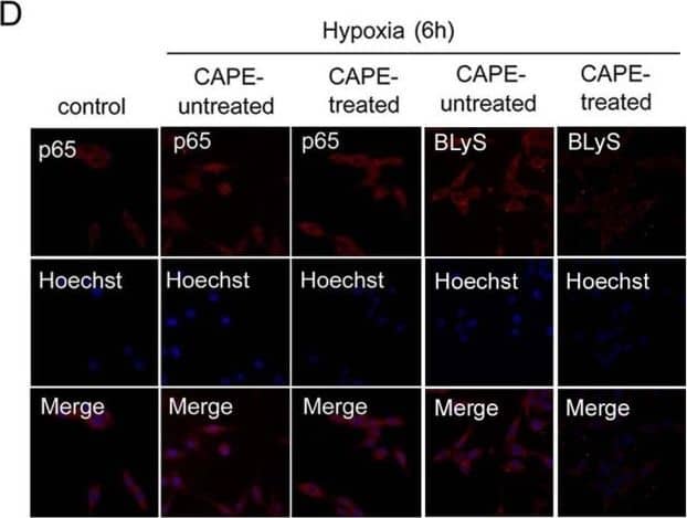
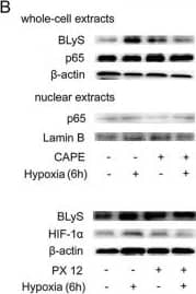
Detection of Human BAFF/BLyS/TNFSF13B by Immunocytochemistry/ Immunofluorescence Role of p65 activation in BLyS up-regulation. (A) HIF-1 alpha and p65 protein levels in MDA-MB-435 in hypoxic conditions for different time points by Western Blotting. (B) CAPE(50 μM)and PX 12 (10 μM) were used to determine the roles of p65 and HIF-1 alpha in the regulation of BLyS expression by Western Blotting. The cells were treated with or without inhibitor in normoxic or hypoxic conditions for 6 h. (C) Effects of CAPE(50 μM)and PX 12 (10 μM) on BLyS promoter activity. Data were average luciferase activities of three independent transfections with ± SD. *, P < 0.05, vs pGL3-Basic/BP. (D) Localization of p65 protein and expression level of BLyS by immunofluorescence. MDA-MB-435 cells were challenged with CAPE (50 μM) for 6 h (original magnification 200 ×). Image collected and cropped by CiteAb from the following open publication (https://jeccr.biomedcentral.com/articles/10.1186/1756-9966-31-31), licensed under a CC-BY license. Not internally tested by R&D Systems.
 View Larger
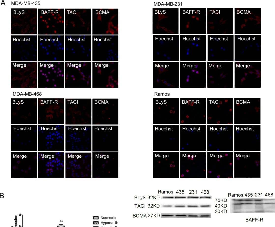
Detection of Human BAFF/BLyS/TNFSF13B by Immunocytochemistry/ Immunofluorescence Expressions of BLyS, TACI, BCMA and BAFF-R in human breast cancer cell lines. (A) BLyS and its three receptors in human breast cancer cell lines MDA-MB-435, MDA-MB-231, MDA-MB-468 and B cell line Ramos by immunofluorescence (original magnification 200 ×) and Western Blotting. (B) The mRNA level of BLyS in the three cell lines were detected by real-time PCR under hypoxia for different time points. Data were means of triplicate samples with ± SD; vs normoxia, *, P < 0.05; **, P < 0.01; ***, P < 0.001. (C) BLyS protein level in MDA-MB-435 cells by Western Blotting analysis. Image collected and cropped by CiteAb from the following open publication (https://jeccr.biomedcentral.com/articles/10.1186/1756-9966-31-31), licensed under a CC-BY license. Not internally tested by R&D Systems.
 View Larger
Detection of Human BAFF/BLyS/TNFSF13B by Western Blot Expressions of BLyS, TACI, BCMA and BAFF-R in human breast cancer cell lines. (A) BLyS and its three receptors in human breast cancer cell lines MDA-MB-435, MDA-MB-231, MDA-MB-468 and B cell line Ramos by immunofluorescence (original magnification 200 ×) and Western Blotting. (B) The mRNA level of BLyS in the three cell lines were detected by real-time PCR under hypoxia for different time points. Data were means of triplicate samples with ± SD; vs normoxia, *, P < 0.05; **, P < 0.01; ***, P < 0.001. (C) BLyS protein level in MDA-MB-435 cells by Western Blotting analysis. Image collected and cropped by CiteAb from the following open publication (https://jeccr.biomedcentral.com/articles/10.1186/1756-9966-31-31), licensed under a CC-BY license. Not internally tested by R&D Systems.
 View Larger
Detection of Human BAFF/BLyS/TNFSF13B by Western Blot Role of p65 activation in BLyS up-regulation. (A) HIF-1 alpha and p65 protein levels in MDA-MB-435 in hypoxic conditions for different time points by Western Blotting. (B) CAPE(50 μM)and PX 12 (10 μM) were used to determine the roles of p65 and HIF-1 alpha in the regulation of BLyS expression by Western Blotting. The cells were treated with or without inhibitor in normoxic or hypoxic conditions for 6 h. (C) Effects of CAPE(50 μM)and PX 12 (10 μM) on BLyS promoter activity. Data were average luciferase activities of three independent transfections with ± SD. *, P < 0.05, vs pGL3-Basic/BP. (D) Localization of p65 protein and expression level of BLyS by immunofluorescence. MDA-MB-435 cells were challenged with CAPE (50 μM) for 6 h (original magnification 200 ×). Image collected and cropped by CiteAb from the following open publication (https://jeccr.biomedcentral.com/articles/10.1186/1756-9966-31-31), licensed under a CC-BY license. Not internally tested by R&D Systems.
Preparation and Storage
- 12 months from date of receipt, -20 to -70 °C as supplied.
- 1 month, 2 to 8 °C under sterile conditions after reconstitution.
- 6 months, -20 to -70 °C under sterile conditions after reconstitution.
Background: BAFF/BLyS/TNFSF13B
BAFF (also known as TALL-1, BLyS, and THANK) is a type II transmembrane glycoprotein belonging to the TNF superfamily and has been designated as TNF superfamily member 13B (TNFSF13B). Human BAFF is a 285 amino acid (aa) protein consisting of a 218 aa extracellular domain, a 21 aa transmembrane region and a 46 aa cytoplasmic tail (1, 2). BAFF has the typical structural characteristics of the TNF superfamily ligands. It is a homotrimeric protein having the structurally conserved motif known as TNF homology domain (3, 4). A higher ordered structure composed of a cluster of trimeric units resembling the structure of a viral capsid has also been reported (4). Human BAFF may be shed from the cell surface by proteolytic cleavage between R133 and Ala134 to yield a soluble form of the protein that is detectable in serum (1, 5). Within the TNF superfamily BAFF shares the highest homology (48%) with APRIL (1). BAFF shares with APRIL the ability to bind to BCMA and TACI and also binds specifically to BAFF receptor (BAFF R, also known as BR3 or TNFSFR13C), which is the principal BAFF receptor (6‑8). All three receptors are type III transmembrane proteins that are expressd in B cells. BAFF and APRIL can form active heteromers that bind to TACI (9). BAFF is expressed in peripheral blood mononuclear cells, in spleen and lymph nodes. Its expression in resting monocytes is up-regulated by IFN-alpha, IFN-beta, LPS and IL-10. BAFF provides critical survival signals to a subset of B cells with intermediate maturation status (T2 B cells) during the immune response (10). BAFF also plays an important role in the development of lymphoid tissue and enhances the survival of activated memory B cells (7, 11). Human and mouse BAFF share 86% aa sequence identity (1).
- Schneider, P. et al. (1999) J. Exp. Med. 189:1747.
- Mukhopadhyay, A. et al. (1999) J. Biol. Chem. 274:15978.
- Karpusas, M. et al. (2002) J. Mol. Biol. 315:1145.
- Liu, Y. et al. (2002) Cell 108:383.
- Cheema, G.S. et al. (2001) Arthr. Rheum. 44:1313.
- Marsters, S.A. et al. (2000) Curr. Biol. 10:785.
- Thompson, J.S. et al. (2001) Science 293:2108.
- Ng, L.G. et al. (2004) J. Immunol. 173:807.
- Roschke, V. et al. (2002) J. Immunol. 169:4314.
- Batten, M. et al. (2000) J. Exp. Med. 192:1453.
- Avery, D.T. et al. (2003) J. Clin. Invest. 112:286.
Product Datasheets
Citations for Human BAFF/BLyS/TNFSF13B Antibody
R&D Systems personnel manually curate a database that contains references using R&D Systems products. The data collected includes not only links to publications in PubMed, but also provides information about sample types, species, and experimental conditions.
15
Citations: Showing 1 - 10
Filter your results:
Filter by:
-
Single-cell and spatial transcriptome analyses reveal tertiary lymphoid structures linked to tumour progression and immunotherapy response in nasopharyngeal carcinoma
Authors: Liu, Y;Ye, SY;He, S;Chi, DM;Wang, XZ;Wen, YF;Ma, D;Nie, RC;Xiang, P;Zhou, Y;Ruan, ZH;Peng, RJ;Luo, CL;Wei, PP;Lin, GW;Zheng, J;Cui, Q;Cai, MY;Yun, JP;Dong, J;Mai, HQ;Xia, X;Bei, JX;
Nature communications
Species: Human
Sample Types: Whole Cells
Applications: Neutralization -
An efficient immunoassay for the B cell help function of SARS-CoV-2-specific memory CD4+ T cells
Authors: Asgar Ansari, Shilpa Sachan, Bimal Prasad Jit, Ashok Sharma, Poonam Coshic, Alessandro Sette et al.
Cell Reports Methods
-
BDNF belongs to the nurse-like cell secretome and supports survival of B chronic lymphocytic leukemia cells
Authors: H Talbot, S Saada, E Barthout, PF Gallet, N Gachard, J Abraham, A Jaccard, D Troutaud, F Lalloué, T Naves, AL Fauchais, MO Jauberteau
Sci Rep, 2020-07-28;10(1):12572.
Species: Human
Sample Types: Whole Cells
Applications: Neutralization -
Monitoring tissue-level remodelling during inflammatory arthritis using a three-dimensional synovium-on-a-chip with non-invasive light scattering biosensing
Authors: Mario Rothbauer, Gregor Höll, Christoph Eilenberger, Sebastian R. A. Kratz, Bilal Farooq, Patrick Schuller et al.
Lab on a Chip
-
BAFF is involved in the pathogenesis of IgA nephropathy by activating the TRAF6/NF?&kappaB signaling pathway in glomerular mesangial cells
Authors: Y Cao, G Lu, X Chen, X Chen, N Guo, W Li
Mol Med Rep, 2019-12-06;21(2):795-805.
Species: Human
Sample Types: Whole Tissue
Applications: IHC -
IFN-? stimulates CpG-induced IL-10 production in B cells via p38 and JNK signalling pathways
Authors: M Imbrechts, K De Samblan, K Fierens, E Brisse, J Vandenhaut, T Mitera, C Libert, I Smets, A Goris, C Wouters, P Matthys
Eur. J. Immunol., 2018-08-01;0(0):.
Species: Human
Sample Types: Whole Cells
Applications: Neutralization -
Plasmacytoid dendritic cells and RNA-containing immune complexes drive expansion of peripheral B cell subsets with an SLE-like phenotype
Authors: O Berggren, N Hagberg, A Alexsson, G Weber, L Rönnblom, ML Eloranta
PLoS ONE, 2017-08-28;12(8):e0183946.
Species: Human
Sample Types: Whole Cells
Applications: Neutralization -
Serum B cell-activating factor (BAFF) level in connective tissue disease associated interstitial lung disease.
Authors: Hamada T, Samukawa T, Kumamoto T, Hatanaka K, Tsukuya G, Yamamoto M, Machida K, Watanabe M, Mizuno K, Higashimoto I, Inoue Y, Inoue H
BMC Pulm Med, 2015-09-30;15(0):110.
Species: Human
Sample Types: Whole Tissue
Applications: IHC-P -
The B Cell-Stimulatory Cytokines BLyS and APRIL Are Elevated in Human Periodontitis and Are Required for B Cell-Dependent Bone Loss in Experimental Murine Periodontitis.
Authors: Abe T, AlSarhan M, Benakanakere M, Maekawa T, Kinane D, Cancro M, Korostoff J, Hajishengallis G
J Immunol, 2015-07-06;195(4):1427-35.
Species: Human
Sample Types: Whole Tissue
Applications: IHC -
The BAFF receptor TACI controls IL-10 production by regulatory B cells and CLL B cells.
Authors: Saulep-Easton D, Vincent F, Quah P, Wei A, Ting S, Croce C, Tam C, Mackay F
Leukemia, 2015-07-03;30(1):163-72.
Species: Human
Sample Types: Whole Cells
Applications: Neutralization -
BlyS is up-regulated by hypoxia and promotes migration of human breast cancer cells.
Authors: Zhu J, Sun L, Lin S
J. Exp. Clin. Cancer Res., 2012-03-31;31(0):31.
Species: Human
Sample Types: Cell Lysates, Whole Cells
Applications: ICC, Western Blot -
Unusual presentation of multiple pathologic bone fractures in a patient with gastric mucosa-associated lymphoid tissue lymphoma.
Authors: Kuo SH, Yen RF, Lin CW, Chen LT, Tien HF, Cheng AL
Ann. Hematol., 2009-08-18;89(4):431-6.
Species: Human
Sample Types: Whole Cells
Applications: ICC -
Essential role of antigen-presenting cell-derived BAFF for antibody responses.
Authors: Bergamin F, Vincent IE, Summerfield A, McCullough KC
Eur. J. Immunol., 2007-11-01;37(11):3122-30.
Species: Porcine
Sample Types: Whole Cells
Applications: ICC -
B-lymphocyte stimulator (BLyS) stimulates immunoglobulin production and malignant B-cell growth in Waldenstrom macroglobulinemia.
Authors: Elsawa SF, Novak AJ, Grote DM, Ziesmer SC, Witzig TE, Kyle RA, Dillon SR, Harder B, Gross JA, Ansell SM
Blood, 2005-11-22;107(7):2882-8.
Species: Human
Sample Types: Whole Tissue
Applications: IHC-P -
Expression profile of BAFF in peripheral blood from patients of IgA nephropathy: Correlation with clinical features and Streptococcus�pyogenes infection
Authors: N Zheng, J Fan, B Wang, D Wang, P Feng, Q Yang, X Yu
Mol Med Rep, 2017-02-10;0(0):.
FAQs
No product specific FAQs exist for this product, however you may
View all Antibody FAQsIsotype Controls
Reconstitution Buffers
Secondary Antibodies
Reviews for Human BAFF/BLyS/TNFSF13B Antibody
Average Rating: 5 (Based on 1 Review)
Have you used Human BAFF/BLyS/TNFSF13B Antibody?
Submit a review and receive an Amazon gift card.
$25/€18/£15/$25CAN/¥75 Yuan/¥2500 Yen for a review with an image
$10/€7/£6/$10 CAD/¥70 Yuan/¥1110 Yen for a review without an image
Filter by:


