Human CD28 Antibody Summary
Asn19-Pro152
Accession # P10747
Customers also Viewed
Applications
Please Note: Optimal dilutions should be determined by each laboratory for each application. General Protocols are available in the Technical Information section on our website.
Scientific Data
 View Larger
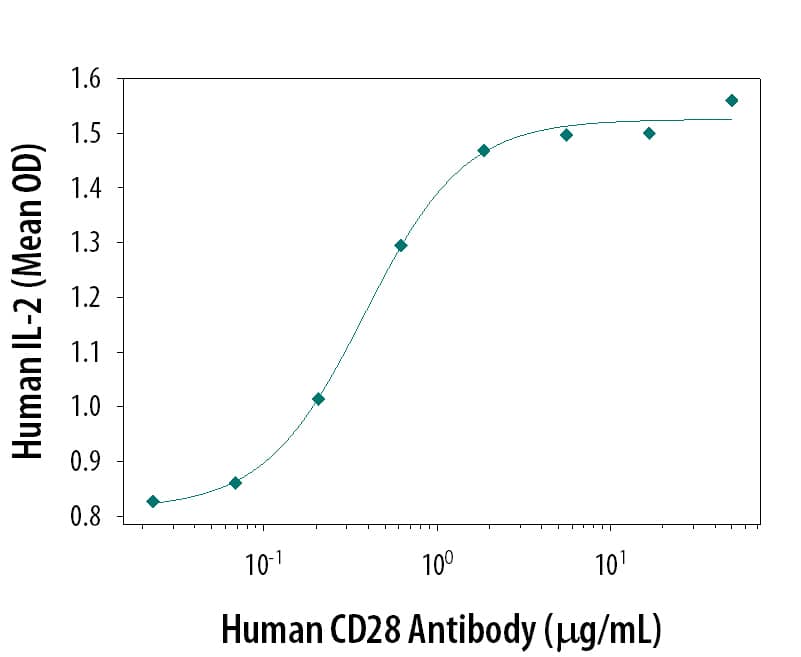
Human CD28 Antibody Enhances IL-2 Secretion in Jurkat Cells. Human CD28 Antigen Affinity-purified Polyclonal Antibody enhances IL-2 secretion in Jurkat human acute T cell leukemia cell line stimulated with 10 ng/mL phorbol myristate acetate (PMA) and 0.5 µM calcium ionophore, in a dose-dependent manner, as measured using the Quantikine Human IL-2 ELISA Kit (Catalog # D2050). The ED50 for this effect is typically 0.3-0.6 µg/mL.
 View Larger
CD28 in Human PBMCs. CD28 was detected in immersion fixed human peripheral blood mononuclear cells (PBMCs) using 10 µg/mL Goat Anti-Human CD28 Antigen Affinity-purified Polyclonal Antibody (Catalog # AF-342-PB) for 3 hours at room temperature. Cells were stained with the NorthernLights™ 557-conjugated Anti-Goat IgG Secondary Antibody (red; Catalog # NL001) and counter-stained with DAPI (blue). View our protocol for Fluorescent ICC Staining of Non-adherent Cells.
 View Larger
CD28 in Human Tonsil. CD28 was detected in immersion fixed paraffin-embedded sections of human tonsil using Goat Anti-Human CD28 Antigen Affinity-purified Polyclonal Antibody (Catalog # AF-342-PB) at 3 µg/mL for 1 hour at room temperature followed by incubation with the Anti-Goat IgG VisUCyte™ HRP Polymer Antibody (VC004). Tissue was stained using DAB (brown) and counterstained with hematoxylin (blue). Specific staining was localized to cell surfaces in lymphocytes. View our protocol for IHC Staining with VisUCyte HRP Polymer Detection Reagents.
Preparation and Storage
- 12 months from date of receipt, -20 to -70 °C as supplied.
- 1 month, 2 to 8 °C under sterile conditions after reconstitution.
- 6 months, -20 to -70 °C under sterile conditions after reconstitution.
Background: CD28
CD28 and CTLA-4, together with their ligands, B7-1 and B7-2, constitute one of the dominant costimulatory pathways that regulate T and B cell responses. CD28 and CTLA-4 are structurally homologous molecules that are members of the immunoglobulin (Ig) gene superfamily. Both CD28 and CTLA-4 are composed of a single Ig V‑like extracellular domain, a transmembrane domain and an intracellular domain. CD28 and CTLA-4 are both expressed on the cell surface as disulfide-linked homodimers or as monomers. The genes encoding these two molecules are closely linked on human chromosome 2 and mouse chromosome 1. Mouse CD28 is expressed constitutively on virtually 100% of mouse T cells and on developing thymocytes. Cell surface expression of mouse CD28 is down-regulated upon ligation of CD28 in the presence of PMA or PHA. In contrast, CTLA-4 is not expressed constitutively but is up-regulated rapidly following T cell activation and CD28 ligation. Cell surface expression of mouse CTLA-4 peaks approximately 48 hours after activation. Although both CTLA-4 and CD28 can bind to the same ligands, CTLA-4 binds to B7-1 and B7-2 with a 20-100 fold higher affinity than CD28. CD28/B7 interaction has been shown to prevent apoptosis of activated T cells via the upregulation of Bcl-xL. CD28 ligation has also been shown to regulate Th1/Th2 differentiation.
- Lenschow, D.J. et al. (1996) Annu. Rev. Immunol. 14:233.
- Hathcock, K.S. and R.J. Hodes (1996) Advances in Immunol. 62:131.
- Ward, S.G. (1996) Biochem. J. 318:361.
Product Datasheets
Citations for Human CD28 Antibody
R&D Systems personnel manually curate a database that contains references using R&D Systems products. The data collected includes not only links to publications in PubMed, but also provides information about sample types, species, and experimental conditions.
8
Citations: Showing 1 - 8
Filter your results:
Filter by:
-
The homodimer interfaces of costimulatory receptors B7 and CD28 control their engagement and pro-inflammatory signaling
Authors: Popugailo, A;Rotfogel, Z;Levy, M;Turgeman, O;Hillman, D;Levy, R;Arad, G;Shpilka, T;Kaempfer, R;
Journal of biomedical science
Species: Mouse
Sample Types: Whole Cells
Applications: Flow Cytometry -
Mesangial Cells Exhibit Features of Antigen-Presenting Cells and Activate CD4+ T Cell Responses
Authors: H Yu, S Cui, Y Mei, Q Li, L Wu, S Duan, G Cai, H Zhu, B Fu, L Zhang, Z Feng, X Chen
J Immunol Res, 2019-06-17;2019(0):2121849.
Species: Human
Sample Types: Whole Cells
Applications: Cell Culture -
Diversification of Bw4 Specificity and Recognition of a Nonclassical MHC Class I Molecule Implicated in Maternal-Fetal Tolerance by Killer Cell Ig-like Receptors of the Rhesus Macaque
Authors: P Banerjee, M Ries, SK Janaka, AG Grandea, R Wiseman, DH O'Connor, TG Golos, DT Evans
J. Immunol., 2018-09-19;0(0):.
Species: Human
Sample Types: Whole Cells
Applications: Flow Cytometry -
Dysregulated CD46 shedding interferes with Th1-contraction in systemic lupus erythematosus
Authors: U Ellinghaus, A Cortini, CL Pinder, GL Friec, C Kemper, TJ Vyse
Eur. J. Immunol., 2017-05-22;0(0):.
Species: Human
Sample Types: Whole Cells
Applications: Functional Assay -
Phenotype, function, and differentiation potential of human monocyte subsets
Authors: LB Boyette, C Macedo, K Hadi, BD Elinoff, JT Walters, B Ramaswami, G Chalasani, JM Taboas, FG Lakkis, DM Metes
PLoS ONE, 2017-04-26;12(4):e0176460.
Species: Human
Sample Types: Whole Cells
Applications: Functional Assay -
Binding of Superantigen Toxins into the CD28 Homodimer Interface Is Essential for Induction of Cytokine Genes That Mediate Lethal Shock
Authors: Gila Arad, Revital Levy, Iris Nasie, Dalia Hillman, Ziv Rotfogel, Uri Barash et al.
PLoS Biology
-
The soluble forms of CD28, CD86 and CTLA-4 constitute possible immunological markers in patients with abdominal aortic aneurysm.
Authors: Sakthivel P, Shively V, Kakoulidou M
J. Intern. Med., 2007-04-01;261(4):399-407.
Species: Human
Sample Types: Plasma
Applications: ELISA Development -
CD28-mediated regulation of multiple myeloma cell proliferation and survival.
Authors: Bahlis NJ, King AM, Kolonias D, Carlson LM, Liu HY, Hussein MA, Terebelo HR, Byrne GE, Levine BL, Boise LH, Lee KP
Blood, 2007-02-20;109(11):5002-10.
Species: Human
Sample Types: Whole Tissue
Applications: IHC
FAQs
No product specific FAQs exist for this product, however you may
View all Antibody FAQsIsotype Controls
Reconstitution Buffers
Secondary Antibodies
Reviews for Human CD28 Antibody
Average Rating: 4 (Based on 2 Reviews)
Have you used Human CD28 Antibody?
Submit a review and receive an Amazon gift card.
$25/€18/£15/$25CAN/¥75 Yuan/¥2500 Yen for a review with an image
$10/€7/£6/$10 CAD/¥70 Yuan/¥1110 Yen for a review without an image
Filter by:


