Human CD40/TNFRSF5 Antibody Summary
Glu21-Arg193
Accession # P25942
Customers also Viewed
Applications
Please Note: Optimal dilutions should be determined by each laboratory for each application. General Protocols are available in the Technical Information section on our website.
Scientific Data
 View Larger
Detection of Human CD40/TNFRSF5 by Western Blot. Western blot shows lysates of Raji human Burkitt's lymphoma cell line and Daudi human Burkitt's lymphoma cell line. PVDF membrane was probed with 2 µg/mL of Goat Anti-Human CD40/TNFRSF5 Antigen Affinity-purified Polyclonal Antibody (Catalog # AF632) followed by HRP-conjugated Anti-Goat IgG Secondary Antibody (HAF017). A specific band was detected for CD40/TNFRSF5 at approximately 40-45 kDa (as indicated). This experiment was conducted under reducing conditions and using Immunoblot Buffer Group 1.
 View Larger
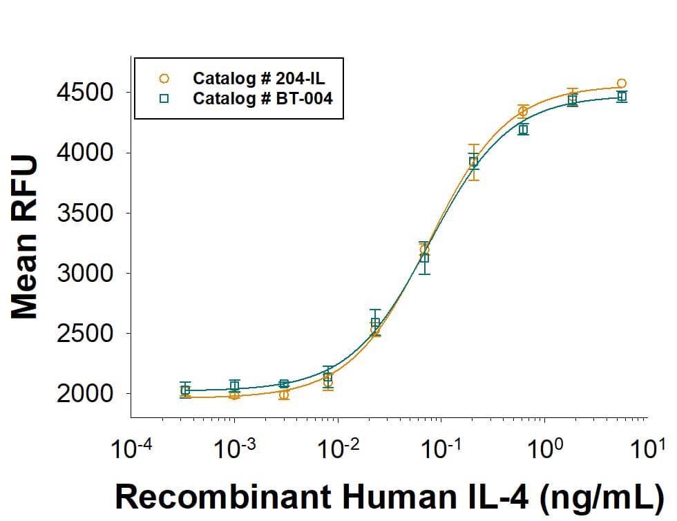
Human CD40/TNFRSF5 Antibody Stimulates Cell Proliferation in Human B Cells. Goat Anti-Human CD40/TNFRSF5 Antigen Affinity-purified Polyclonal Antibody (Catalog # AF632) stimulates human B cell proliferation in the presence of Recombinant Human IL-4 (204-IL) in a dose-dependent manner, as measured by Resazurin (AR002). The ED50 for this effect is typically 3-12 ng/mL
 View Larger
Detection of Human CD40/TNFRSF5 by Simple WesternTM. Simple Western lane view shows lysates of Raji human Burkitt's lymphoma cell line and Daudi human Burkitt's lymphoma cell line, loaded at 0.2 mg/mL. A specific band was detected for CD40/TNFRSF5 at approximately 57 kDa (as indicated) using 50 µg/mL of Goat Anti-Human CD40/TNFRSF5 Antigen Affinity-purified Polyclonal Antibody (Catalog # AF632). This experiment was conducted under reducing conditions and using the 12-230 kDa separation system.
 View Larger
Detection of Fungus CD40/TNFRSF5 by Western Blot SDS–PAGE and Western blot analysis of recombinant CD40-N expressed in P. pastoris. a A 20-μL sample of supernatant was loaded onto a 12 % SDS–PAGE gel and stained with Coomassie brilliant blue G-250. All of the SDS-PAGE experiments were performed under the same conditions. Samples were collected at 12-h intervals for 84 h. The sizes of molecular weight markers (kDa) are shown (M), and recombinant CD40-N protein is indicated with an arrow. b Western blot analysis of human Flag-tagged CD40-N (not undergoing codon optimization) and CD40-N-S (with signal sequence and not undergoing codon optimization) expressed in HEK293T cells. pCDNA3.3-CD40-N and pCDNA3.3-CD40-N-S were introduced into HEK293T cells, and the cell lysates were harvested after 48 h. A 10-μl sample was loaded onto a 12 % SDS–PAGE gel. Anti-Flag, anti-CD40-N and anti-GAPDH antibodies were used to detect the proteins. c Western blot analysis of proteins at different times in P. pastoris (0, 12, 24, 36, 48, 60, 72, and 84 h). P indicates the positive control (CD40-N expressed in HEK293T cells). Image collected and cropped by CiteAb from the following open publication (https://pubmed.ncbi.nlm.nih.gov/26809818), licensed under a CC-BY license. Not internally tested by R&D Systems.
 View Larger
Detection of Yeast CD40/TNFRSF5 by Western Blot SDS–PAGE and Western blot analysis of recombinant CD40-N expressed in P. pastoris. a A 20-μL sample of supernatant was loaded onto a 12 % SDS–PAGE gel and stained with Coomassie brilliant blue G-250. All of the SDS-PAGE experiments were performed under the same conditions. Samples were collected at 12-h intervals for 84 h. The sizes of molecular weight markers (kDa) are shown (M), and recombinant CD40-N protein is indicated with an arrow. b Western blot analysis of human Flag-tagged CD40-N (not undergoing codon optimization) and CD40-N-S (with signal sequence and not undergoing codon optimization) expressed in HEK293T cells. pCDNA3.3-CD40-N and pCDNA3.3-CD40-N-S were introduced into HEK293T cells, and the cell lysates were harvested after 48 h. A 10-μl sample was loaded onto a 12 % SDS–PAGE gel. Anti-Flag, anti-CD40-N and anti-GAPDH antibodies were used to detect the proteins. c Western blot analysis of proteins at different times in P. pastoris (0, 12, 24, 36, 48, 60, 72, and 84 h). P indicates the positive control (CD40-N expressed in HEK293T cells) Image collected and cropped by CiteAb from the following open publication (https://pubmed.ncbi.nlm.nih.gov/26809818), licensed under a CC-BY license. Not internally tested by R&D Systems.
Preparation and Storage
- 12 months from date of receipt, -20 to -70 °C as supplied.
- 1 month, 2 to 8 °C under sterile conditions after reconstitution.
- 6 months, -20 to -70 °C under sterile conditions after reconstitution.
Background: CD40/TNFRSF5
CD40 is a type I transmembrane glycoprotein belonging to the TNF receptor superfamily. The mature hCD40 consists of a 172 amino acid (aa) extracellular domain, a 22 aa transmembrane region and a 62 aa cytoplasmic domain (1). Human and mouse CD40 share 62% aa identity. CD40 is expressed in B cells, follicular dendritic cells, dendritic cells, activated monocytes, macrophages, endothelial cells, vascular smooth muscle cells, and several tumor cell lines (2). The extracellular domain has the cysteine-rich repeat regions, which are characteristic for many of the receptors of the TNF superfamily. Interaction of CD40 with its ligand, CD40L, leads to aggregation of CD40 molecules, which in turn interact with cytoplasmic components to initiate signaling pathways. Early studies on the CD40-CD40L system revealed its role in humoral immunity. Interaction between CD40L on T cells and CD40 on B cells stimulated B cell proliferation and provided the signal for immunoglobulin isotype switching (3). Mutations in the CD40L gene, which resulted in a CD40L molecule unable to interact with CD40, are responsible for the hyper-IgM syndrome (4). Cross-linking of CD40 with antibodies or by CD40 binding to CD40L produces cell type-specific responses which include costimulation and induction of proliferation, induction of cytokine production, rescue from apoptosis, and upregulation of adhesion molecules (5). Some of the early events of intracellular signaling by the CD40-CD40L system include the association of the CD40 with TRAFs and the activation of various kinases (6 - 8).
- Torres, R.M. and E.A. Clark (1992) J. Immunol. 148:620.
- Schonbeck, U. et al. (1997) J. Biol. Chem. 272:19569.
- Armitage, R.J. et al. (1993) J. Immunol. 150:3671.
- Callard, R.E. et al. (1993) Immunol. Today 14:559.
- Stout, R.D. and J. Suttles (1996) Immunol. Today 17:487.
- Pullen, S.S. et al. (1999) Biochemistry 38:10168.
- Faris, M. et al. (1994) J. Exp. Med. 179:1923.
- Hanissian, S.H. and R.S Geha (1997) Immunity 6:379.
Product Datasheets
Citations for Human CD40/TNFRSF5 Antibody
R&D Systems personnel manually curate a database that contains references using R&D Systems products. The data collected includes not only links to publications in PubMed, but also provides information about sample types, species, and experimental conditions.
7
Citations: Showing 1 - 7
Filter your results:
Filter by:
-
Children with oligoarticular juvenile idiopathic arthritis have skewed synovial monocyte polarization pattern with functional impairment-a distinct inflammatory pattern for oligoarticular juvenile arthritis
Authors: T Schmidt, E Berthold, S Arve-Butle, B Gullstrand, A Mossberg, F Kahn, AA Bengtsson, B Månsson, R Kahn
Arthritis Res. Ther., 2020-08-12;22(1):186.
Species: Human
Sample Types: Whole Tissue
Applications: IHC -
Expression, purification and biological characterization of the extracellular domain of CD40 from Pichia pastoris.
Authors: Zhan Y, Wei Y, Chen P, Zhang H, Liu D, Zhang J, Liu R, Chen R, Zhang J, Mo W, Zhang X
BMC Biotechnol, 2016-01-25;16(1):8.
Species: Human
Sample Types: Cell Lysates
Applications: Western Blot -
CD19 and CD32b differentially regulate human B cell responsiveness.
Authors: Karnell J, Dimasi N, Karnell F, Fleming R, Kuta E, Wilson M, Wu H, Gao C, Herbst R, Ettinger R
J Immunol, 2014-01-17;192(4):1480-90.
Species: Human
Sample Types: Whole Cells
Applications: Neutralization -
CD40.FasL inhibits human T cells: evidence for an auto-inhibitory loop-back mechanism.
Authors: Dranitzki-Elhalel M, Huang JH, Sasson M, Rachmilewitz J, Parnas M, Tykocinski ML
Int. Immunol., 2007-02-20;19(4):355-63.
Species: Human
Sample Types: Cell Lysates, Whole Cells
Applications: Western Blot -
Disruption of CD40/CD40 ligand interaction with cleavage of CD40 on human gingival fibroblasts by human leukocyte elastase resulting in down-regulation of chemokine production.
Authors: Nemoto E, Tada H, Shimauchi H
J. Leukoc. Biol., 2002-09-01;72(3):538-45.
Species: Human
Sample Types: Cell Lysates
Applications: Western Blot -
A PDGFRB- and CD40-targeting bispecific AffiMab induces stroma-targeted immune cell activation
Authors: Alessandro Mega, Aman Mebrahtu, Gustav Aniander, Eva Ryer, Annette Sköld, Anna Sandegren et al.
mAbs
-
Bispecific antibody target pair discovery by high-throughput phenotypic screening using in vitro combinatorial Fab libraries
Authors: Pallavi Bhatta, Kevin D. Whale, Amy K. Sawtell, Clare L. Thompson, Stephen E. Rapecki, David A. Cook et al.
mAbs
FAQs
No product specific FAQs exist for this product, however you may
View all Antibody FAQsIsotype Controls
Reconstitution Buffers
Secondary Antibodies
Reviews for Human CD40/TNFRSF5 Antibody
Average Rating: 5 (Based on 1 Review)
Have you used Human CD40/TNFRSF5 Antibody?
Submit a review and receive an Amazon gift card.
$25/€18/£15/$25CAN/¥75 Yuan/¥2500 Yen for a review with an image
$10/€7/£6/$10 CAD/¥70 Yuan/¥1110 Yen for a review without an image
Filter by:


